. 2025 Apr 5;26(7):3416.
doi: 10.3390/ijms26073416.

Characterization of Isolated Human Astrocytes from Aging Brain

Affiliations

Characterization of Isolated Human Astrocytes from Aging Brain

Geidy E Serrano et al. Int J Mol Sci. .

Abstract

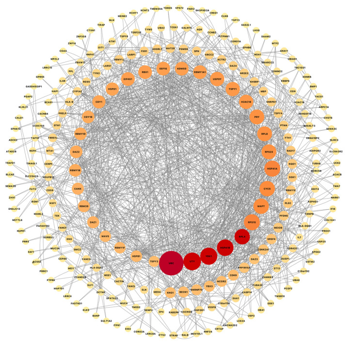
Astrocytes have multiple crucial roles, including maintaining brain homeostasis and synaptic function, performing phagocytic clearance, and responding to injury and repair. It has been suggested that astrocyte performance is progressively impaired with aging, leading to imbalances in the brain's internal milieu that eventually impact neuronal function and lead to neurodegeneration. Until now, most evidence of astrocytic dysfunction in aging has come from experiments done with whole tissue homogenates, astrocytes collected by laser capture, or cell cultures derived from animal models or cell lines. In this study, we used postmortem-derived whole cells sorted with anti-GFAP antibodies to compare the unbiased, whole-transcriptomes of human astrocytes from control, older non-impaired individuals and subjects with different neurodegenerative diseases, such as Parkinson's disease (PD), Alzheimer's disease (ADD), and progressive supranuclear palsy (PSP). We found hundreds of dysregulated genes between disease and control astrocytes. In addition, we identified numerous genes shared between these common neurodegenerative disorders that are similarly dysregulated; in particular, UBC a gene for ubiquitin, which is a protein integral to cellular homeostasis and critically important in regulating function and outcomes of proteins under cellular stress, was upregulated in PSP, PD, and ADD when compared to control.

Keywords: Alzheimer’s disease; Parkinson’s disease; autophagy; neurodegeneration; progressive supranuclear palsy; ubiquitinization; whole-cell-suspension.


Conflict of interest statement

The authors declare no conflicts of interest.

Figures

Figure 1
Figure 1
GFAP immunostains for quality assessments. (A) Whole-cell-dissociated-suspensions pellets were stained for GFAP before sorting to do quality assessments (Scale bar = 20 µm). and (B) after sorting to confirm astrocyte enrichment in our preparations. Example of whole astrocyte in suspension. (Scale bar = 10 µm).
Figure 2
Figure 2
Frontal cortex tissue adjacent to the sample collected for whole-cell-dissociated-suspensions stained for GFAP showed an increased number of reactive astrocytes in PSP cases (D,E), especially when compared to (A) controls, (B) ADD, and (C) PD. (Scale bar on (AE) = 50 µm). (F) The area occupied, measured using Image J version 1.48, showed a significant increase of GFAP positive reactive astrocytes compared to all other diagnostic groups (* p < 0.05 Tukey’s multiple comparisons test; ns = non statistically significant).
Figure 3
Figure 3
Volcano Plots show dysregulated genes obtained from differential expression analysis comparing gene expression from each diseased group to CN, as well as disease comparison. (A) PSP cases had the largest number of dysregulated genes when compared to CN with a total of 1933 upregulated genes (red) and 35 downregulated genes (blue). (B) When PSP was compared to ADD, we observed a total of 89 genes dysregulated. (C) and when compared to PD 243 genes. (D) ADD comparison to CN resulted in 754 genes dysregulated. (E) and only 4 genes when compared to PD. (F) While the PD comparison to CN resulted in 1150 dysregulated genes. (G) 135 when PD was compared to ILBD. (H) ILBD only had one gene dysregulated when compared to CN, and (I) all disease cases showed a total of 501 genes when all cases were combined and compared to CN.
Figure 4
Figure 4
Pathway enrichment analysis was obtained from differential expression analysis comparing gene expression from each diseased group to CN, as well as disease comparison. Normalized enrichment score (NES) of genes associated with specific pathways (X axis) either downregulated (negative values) or upregulated (positive values). The size of each bubble correlates to the number of reads related to each pathway, with larger bubbles representing a greater number of genes, while the color correlates to greater or more statistically significant dysregulation, with red representing a stronger dysregulation than blue. (A) PSP and CN cases comparison showed an enrichment of dysregulated genes associated with multiple biological pathways, some of the most important involving synaptic pathways, chemokine signaling, protein modification processes and proteolysis, while compared to AD, we also observed dysregulation of cellular senescence, cytokine-cytokine receptor, Inositol phosphate metabolism, and other cellular processes and interactions (B). (C) ADD shows dysregulation of synapses, proteolysis, and signaling pathways when compared to CN. (D) Compared to CN, PD showed dysregulation of the translocation of olfactory receptors, synapses, synaptic vesicle cycle, and neurotrophic signaling pathways.
Figure 5
Figure 5
Cytoscape was used to visualize gene connectivity. Highly connected genes appear larger and green, while less connected genes are shown smaller and in yellow, indicating fewer interactions. The protein–protein interactions network from a list of 691 common dysregulated genes for each disease vs. CN comparison shows 541 nodes and 4952 edges; the overlapping analysis identified five common hub genes (dark green; ACTB, EGFR, CALM3, HSP90AB1, and UBC).
Figure 6
Figure 6
Cytoscape was used to visualize gene connectivity. Highly connected genes appear larger and red, while less connected genes are shown smaller and light orange, indicating fewer interactions. Protein–protein interactions network from a list of 501 common genes from a differential expression analysis combining all disease cases and comparing them to all non-symptomatic cases shows 268 nodes and 1124 edges, while the overlapping analysis identified five common hub genes (red; UBC, YBX1, UTY, RPL4, and HSPA1B).
Figure 7
Figure 7
The list of common hub genes affected in astrocytes from multiple neurodegenerative diseases. We used the network and hub genes identified in Figure 5 and Figure 6 and identified and validated UBC, RPL4, and HSPA1A as important hub genes whose networks seem to be commonly affected in PSP, ADD, and PD astrocytes.

Similar articles

References

    1. Booth H.D.E., Hirst W.D., Wade-Martins R. The Role of Astrocyte Dysfunction in Parkinson’s Disease Pathogenesis. Trends Neurosci. 2017;40:358–370. doi: 10.1016/j.tins.2017.04.001. - DOI - PMC - PubMed
    1. Sorrentino Z.A., Giasson B.I., Chakrabarty P. α-Synuclein and astrocytes: Tracing the pathways from homeostasis to neurodegeneration in Lewy body disease. Acta Neuropathol. 2019;138:1–21. doi: 10.1007/s00401-019-01977-2. - DOI - PMC - PubMed
    1. Fan L.Y., Yang J., Li M.L., Liu R.Y., Kong Y., Duan S.Y., Guo G.Y., Yang J.H., Xu Y.M. Single-nucleus transcriptional profiling uncovers the reprogrammed metabolism of astrocytes in Alzheimer’s disease. Front. Mol. Neurosci. 2023;16:1136398. doi: 10.3389/fnmol.2023.1136398. - DOI - PMC - PubMed
    1. Miyazaki I., Asanuma M. Neuron-Astrocyte Interactions in Parkinson’s Disease. Cells. 2020;9:2623. doi: 10.3390/cells9122623. - DOI - PMC - PubMed
    1. Benarroch E.E. Astrocyte signaling and synaptic homeostasis: I: Membrane channels, transporters, and receptors in astrocytes. Neurology. 2016;87:324–330. doi: 10.1212/WNL.0000000000002875. - DOI - PubMed

MeSH terms

LinkOut - more resources