Review
. 2025 Jul;37(27):e2420114.
doi: 10.1002/adma.202420114. Epub 2025 Apr 17.

Materials Advances in Devices for Heart Disease Interventions

Affiliations
Review

Materials Advances in Devices for Heart Disease Interventions

Gagan K Jalandhra et al. Adv Mater. 2025 Jul.

Abstract

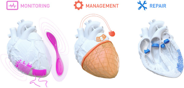
Heart disease encompasses a range of conditions that affect the heart, including coronary artery disease, arrhythmias, congenital heart defects, heart valve disease, and conditions that affect the heart muscle. Intervention strategies can be categorized according to when they are administered and include: 1) Monitoring cardiac function using sensor technology to inform diagnosis and treatment, 2) Managing symptoms by restoring cardiac output, electrophysiology, and hemodynamics, and often serving as bridge-to-recovery or bridge-to-transplantation strategies, and 3) Repairing damaged tissue, including myocardium and heart valves, when management strategies are insufficient. Each intervention approach and technology require specific material properties to function optimally, relying on materials that support their action and interface with the body, with new technologies increasingly depending on advances in materials science and engineering. This review explores material properties and requirements driving innovation in advanced intervention strategies for heart disease and highlights key examples of recent progress in the field driven by advances in materials research.

Keywords: biomaterials; cardiovascular disease; devices; heart disease; tissue engineering.


Conflict of interest statement

The authors declare no conflict of interest.

Figures

Figure 1
Figure 1
Monitoring, Management, and Repair represent three key stages in heart disease intervention. (Left) Visual representation of technologies discussed in this review for monitoring of cardiovascular function and heart disease: wearable devices—worn externally—which interface with skin, and implantable sensors (i.e., epicardial bioelectronics)—which directly interface with cardiac tissue. Monitoring is conducted wirelessly or via cables. (Center) Technologies discussed in this review for management of heart disease symptoms. Devices pictured: a soft robotic ventricular assist sleeve, traditional pacemaker, and cardiac stimulation patch. (Right) Repair strategies discussed in this review of commonly affected cardiac tissue, including myocardium (multifunctional hydrogel biomaterials) and valves (bioprosthetic or tissue‐engineered).
Figure 2
Figure 2
Wearable ECG electrodes on soft polymeric substrates for conformal adhesion to skin. A) The Zio patch is an FDA‐approved wearable device for single‐lead ECGs which allows continuous monitoring of cardiac function. Reproduced with permission.[ 8 ] Copyright, 2019, Springer Nature Limited. (Credit: iRhythm Technologies). B) Epidermal electronic system using a gold filamentary serpentine configuration. Reproduced with permission.[ 30 ] Copyright, 2013, WILEY‐VCH. C) Schematic illustrating the general process of flexible electrode formation including: coating a flexible polymer (e.g., PMMA) onto a rigid substrate (e.g., Si), evaporating and patterning (typically via lithography) of desired metal, and finally encapsulation of noncontacting areas. Flexible electrodes are typically characterized to evaluate their (i) electrochemical, (ii) electrical, and (iii) mechanical performance to ensure reliable operation in situ. D) Stretchable electrodes made using nanowires and PDMS. Reproduced with permission.[ 18 ] Copyright, 2018, RSC Publishing. E) Ultraconformable temporary tattoo devices based on inkjet‐printed PEDOT:PSS electrodes and gold interconnects (scale bar: 2 cm) (i) Assembled circular electrode transferred on the arm; (ii) SEM images of a conformal contact between the E‐tattoo with a silicon substrate. (scale bar: 400 µm). Reproduced with permission.[ 26 ] Copyright, 2018, WILEY‐VCH. F) SEM images of gecko‐inspired pillar structures to enhance adhesion between devices and skin (scale bar: 20 µm). Reproduced with permission.[ 29 ] Copyright, 2016, American Chemical Society.
Figure 3
Figure 3
Wearable mechano‐acoustic sensors. A) Illustration of basic principles of the mechanisms behind commonly utilized mechano‐acoustic sensors. i) Piezoresistive: applied pressure on a diaphragm causes tensile strain in a piezoresistive material. The tensile strain is directly proportional to the change in resistance. ii) Piezoelectric: application of pressure to piezoelectric crystals or ceramics leads to charge generation across the face of the material. iii) Capacitive: applied pressure changes the distance between two parallel conductive plates which changes the capacitance of the circuit. B) A capacitive sensor using helical Ag electrodes printed on polymeric fiber. Right: SEM images of the Ag electrode printed on the elastic TPU fiber. Reproduced with permission.[ 32 ] Copyright, 2023, Chi Zhang et al. C) Integration of acceleration sensors, MEMS microphone and Bluetooth low‐energy (BLE) for wireless, wide‐band‐width acoustic sensors. Right: The device was attached and tested on a 15‐month neonate to monitor their body sounds. Reproduced with permission.[ 35 ] Copyright, 2023, Springer Nature Limited. D) Wearable ultrasound imaging device using a piezoelectric array and liquid metal interconnect. Reproduced with permission.[ 36 ] Copyright, 2023, Hongjie Hu et al.
Figure 4
Figure 4
Oxygen saturation measurement using wearable optical sensors. A) The basic principle of photoplethysmography. Devices generally consist of a light‐source (LED) and a photodetector. Changes in light either reflected (configuration on the left) or transmitted (configuration on the right) correspond to changes in the volume of blood in vessels during cardiac cycles. B) Pulse Oximetry in Ambient Light using Organic Optoelectronics. Inset: image of the photodiode. Reproduced with permission.[ 46 ] Copyright, 2020, WILEY‐VCH. B) Wearable QD‐LED optical sensors using wavy, pretrained LED. Top left: SEM image of wavy structure. Top right: The LED retains stable performance under mechanical strain. Bottom, demonstration of pulse measurement. Reproduced with permission.[ 47 ] Copyright, 2017, American Chemical Society.
Figure 5
Figure 5
Implanted electronic patches. A) Multifunctional sensors and electrodes for endocardial balloon catheter. Reproduced with permission.[ 53 ] Copyright, 2020, Springer Nature Limited. (scale bars: (i),(ii) 500 µm, (iii) 500 µm (left), and 100 µm (right)). B) i) Design process for 3D multifunctional integumentary membrane (3D‐MIMS) including imaging and sectioning of cardiac tissue for topography matching. ii) Photograph of a device on Langendorff‐perfused rabbit heart. Rproduced with permission.[ 59 ] Copyright, 2014, Springer Nature Limited. (scale bars: 6 mm). C) An epicardial bioelectronic device for electrocortical mapping based entirely on rubbery elements. Reproduced with permission.[ 51 ] Copyright, 2020, Springer Nature Limited. D) Simultaneous optical pacing and electrical recording using flexible printed circuits integrated with micro‐LEDs and Pt microelectrodes. Reproduced with permission.[ 67 ] Copyright, 2022, The American Association for the Advancement of Science.
Figure 6
Figure 6
Implanted mechanical sensors for hemodynamic monitoring. A) A photograph of the CardioMEMS device used for wireless blood pressure measurement. Reproduced with permission.[ 78 ] Copyright, 2020, Jacob Abraham et al. B) 3D printed stent integrated with pressure sensor and inductive coupling antenna. Reproduced with permission.[ 77 ] Copyright, 2022, The American Association for the Advancement of Science. C) Multimodal implanted sensors with a BLE module, capable of measuring blood flow rate, blood pressure, and temperature. Reproduced with permission.[ 52 ] Copyright, 2023, Springer Nature Limited.
Figure 7
Figure 7
Overview of traditional VAD operation, complications and surface treatments. A) Implantation and operation of a traditional centrifugal VAD. B) Implantation and operation of an axial VAD. C) Locations of potential pump thrombus development. D) A range of surface treatments for VAD pumps to reduce the risk of pump thrombosis: Diamond like carbon (DLC), 2‐methacryloyloxyethyl phosphorylcholine (MPC), heparin, and surface texturing.
Figure 8
Figure 8
Soft Active Ventricular Assist Devices. A) The AudiCor reBEAT device consisting of inflatable balloons. A driveline connects the implanted device to an external portable drive unit. Reproduced with permission.[ 98 ] Copyright, 2022, The American Association for Thoracic Surgery. B) i) Soft VAD robotic sleeve based on McKibbon artificial muscle fibers arranged circumferentially and axially to allow compression and twisting motion. ii) Circumferential (top) and twisting (bottom) actuators on a porcine heart cadaver. Reproduced with permission.[ 102 ] Copyright, 2017, The American Association for the Advancement of Science. C) Electrothermally‐actuated artificial muscle fibers based on silver‐coated nylon. (scale bar: 1 mm) Reproduced with permission.[ 103 ] Copyright, 2021, Wiley‐VCH. D) VAD sleeve based on hydraulically actuated artificial muscle fibers. ii) Arrangement of artificial muscle fibers to induce contraction and twisting motion. iii) Artificial pericardium deployed to ensure force transfer from the device to the underlying muscle. Reproduced with permission.[ 89 ] Copyright, 2023, Wiley‐VCH.
Figure 9
Figure 9
Advances in biocompatible skins A–C) and actuation mechanisms D,E) for catheter‐based endovascular approaches. A) Ultrathin hydrogel coatings on medical catheter surfaces mitigating bacterial adhesion and thrombosis. Reproduced with permission.[ 111 ] Copyright, 2020, Wiley‐VCH. B) PSHV‐N+ Si coatings on intravascular catheters’ surface for antibacterial, antiadhesion, and low‐friction functions wit h stability. Reproduced with permission.[ 133 ] Copyright, 2022, Wiley‐VCH. C) Heparin coatings central venous catheters’ surface with robust, antibacterial, and antithrombotic properties. Reproduced with permission.[ 109 ] Copyright, 2024, Springer Nature Limited. D) Schematic illustration of a ferromagnetic soft continuum robotic catheter with programmed magnetic polarities, resulting from NdFeB/PDMS composite, and a hydrogel skin (left). Demonstration of navigating through a 3D tortuous and narrow phantom (right). Reproduced with permission.[ 125 ] Copyright, 2019, The American Association for the Advancement of Science. E) i) Design and working principle of a 900‐µm‐diameter end‐effector of a hydraulically steerable catheter. ii) Cross‐section images with the components annotated. Reproduced with permission.[ 114 ] Copyright, 2021, The American Association for the Advancement of Science.
Figure 10
Figure 10
Different types of pacemakers. A) Transvenous pacemaker. B) Leadless pacemaker. C) Epicardial pacemaker.
Figure 11
Figure 11
Advances in cardiac patch biomaterials for myocardial tissue engineering. A) Paintable hydrogel based on decellularized porcine cardiac ECM combined with hyaluronic acid modified with tyramine for the formation of cardiac patches in situ. Reproduced with permission.[ 192 ] Copyright, 2024, Wiley‐VCH. B) i) Visualization of the fiber orientation and changes in angle (left) in the left ventricle from the epicardium to the endocardium. iii) 3D stretchable architecture provides dynamic stretchability with the deforming heart throughout systole and diastole. Reproduced with permission.[ 200 ] Copyright, 2020, The American Association for the Advancement of Science. C) Intrapericardial delivery of biomaterial‐assisted exosome delivery for MI repair in a mouse model. Reproduced with permission.[ 188 ] Copyright, 2021, Dashuai Zhu et al. D) Curcumin nanoparticle‐loaded gelatin nanoparticles (GelB‐Cur NPs) and recombinant human collagen III (rhCol III) are loaded into a carboxymethyl chitosan and oxydextrin scaffold for rapid curcumin and sustained rhCol III release to treat MI. Reproduced with permission.[ 201 ] Copyright, 2023, Elsevier B.V.
Figure 12
Figure 12
Material and biofabrication advances for polymeric heart valves. A) Biomimetic, tri‐layered valves using polycarbonate‐based polyurethane (PCU) porous foam covered by electrospun polycaprolactone (PCL)‐enhanced PCU films. Leaflet‐substitute material demonstrates improved biostability, durability, flexibility, and anticalcification potential compared to most commercial patches. Reproduced with permission.[ 246 ] Copyright, 2022, Elsevier Ltd. B) Bioinspired silicone heart valves using direct ink writing. A heart‐valve‐shaped mandrel is spray‐coated with different stiffness silicone to create bilayered leaflets. Biomimetic fiber supports, inter‐leaflet edges and a stent‐like frame are printed onto the leaflets. Finite element analysis and accelerated wear testing demonstrate superior material durability and high fatigue resistance of the fiber‐reinforced leaflets. Reproduced with permission.[ 247 ] Copyright, 2019, Elsevier Inc. C) Stereolithography (SLA) printed heterogenous polymeric heart valve from a polyacrylamide‐polyacrylic acid (PAAm‐PAA) hydrogel and strengthened with carboxyl‐Fe3+ complexes. Printed hydrogel skeleton is injected molded with a soft hydrogel PAAm matrix to form a composite to allow the polymer skeleton and matrix to entangle topologically. Hydrogel valve remains intact after 10 000 cycles in a hemodynamic test system. Reproduced with permission.[ 248 ] Copyright, 2021, Elsevier Inc. D) Whole heart valve via SLA printing of urea‐based poly(N‐acryloylsemicarbazide‐co‐acrylamide) (P(NASC‐co‐Aam)) hydrogel ink and surface functionalization with heparin‐like sodium polystyrene sulfonate (PSS) chains via reversible addition‐fragmentation chain transfer (RAFT) polymerization to reduce thrombogenicity and hemolysis. Reproduced with permission.[ 249 ] Copyright, 2022, American Chemical Society.
Figure 13
Figure 13
Material and biofabrication advances for in situ tissue‐engineered heart valves. A) (Left) Bis‐urea‐modified polycarbonate (PC‐BU) elastomer electrospun onto polyetheretherketone (PEEK) frame to form a porous, microfibrous heart valve. (Center) Implanted acellular scaffold triggers sequential recruitment of inflammatory, progenitor, and ECM‐producing cells, driving host tissue remodeling and regeneration to form a living, autologous heart valve. (Right) Explanted after 12‐months of implantation in the pulmonary position in sheep shows neo‐matrix formation in leaflets. Reproduced with permission.[ 261 ] Copyright, 2017, Elsevier Ltd. B) Melt‐electro‐writing (MEW) produces serpentine architecture poly(caprolactone) (PCL) tubular scaffold. Resulting MEW‐PCL macroporous scaffold injection‐molded with elastin‐like recombinamer (ELR) hydrogel for favorable cellular infiltration and hemocompatibility. Reproduced with permission.[ 268 ] Copyright, 2022, Wiley‐VCH. C) Soft and elastic PCL fibrous heart valve (FibraValve) fabricated using focused rotary jet printing (PRJS) manufactured in less than 10 min by increasing the rate of fiber deposition on a rotating mandrel. Biomimetic fibrous valve is interfaced with expandable stent for transcatheter delivery, readily supporting initial cellular adhesion and infiltration, both in vitro and in large‐animal model. Reproduced with permission.[ 270 ] Copyright, 2023 Elsevier Inc. D) Cell‐free direct ink writing printed aligned PCL fiber scaffold, replicating the orientation and structural rigidity in the fibrosa layer of native leaflets. The fiber‐reinforcing PCL layer is molded with a cell‐laden 3D‐printed gelatin methacrylate and polyethylene glycol diacrylate (GelMA/PEGDA) bioink layer, encapsulating valvular interstitial‐like cells, to develop a multilayered leaflet capable of repair and remodeling. Reproduced with permission (left).[ 271 ] Copyright, 2020, Elsevier Ltd. Reproduced with permission (center and right).[ 273 ] Copyright, 2024, Wiley‐VCH GmbH.
Figure 14
Figure 14
Summary of future directions in material advancements in devices for heart disease interventions. As discussed in Future directions, key areas of advancement for improved materials design for heart disease interventions are additive manufacture and artificial intelligence (including machine learning). Advances in these fields reduce the cost and time taken to develop new and improved materials for use in heart disease interventions, leading to improved outcomes across the three themes (monitoring, management, and repair) discussed in this paper.

Similar articles

Cited by

References

    1. Martin S. S., Aday A. W., Almarzooq Z. I., Anderson C. A. M., Arora P., Avery C. L., Baker‐Smith C. M., Barone Gibbs B., Beaton A. Z., Boehme A. K., Commodore‐Mensah Y., Currie M. E., Elkind M. S. V., Evenson K. R., Generoso G., Heard D. G., Hiremath S., Johansen M. C., Kalani R., Kazi D. S., Ko D., Liu J., Magnani J. W., Michos E. D., Mussolino M. E., Navaneethan S. D., Parikh N. I., Perman S. M., Poudel R., Rezk‐Hanna M., et al., Circulation 2024, 149, 347.
    1. Townsend N., Kazakiewicz D., Lucy Wright F., Timmis A., Huculeci R., Torbica A., Gale C. P., Achenbach S., Weidinger F., Vardas P., Nat. Rev. Cardiol. 2022, 19, 133. - PubMed
    1. Mirzajani H., Kraft M., ACS Sens. 2024, 9, 4328. - PubMed
    1. Galli A., Ambrosini F., Lombardi F., Arrhythmias Electrophysiol. Rev. 2016, 5, 136. - PMC - PubMed
    1. Rothman S. A., Laughlin J. C., Seltzer J., Walia J. S., Baman R. I., Siouffi S. Y., Sangrigoli R. M., Kowey P. R., J. Cardiovasc. Electrophysiol. 2007, 18, 241. - PubMed

Substances

LinkOut - more resources