

A Novel Approach to Fabricate Early Keratoconus Phantom Models
- PMID: 40244577
- PMCID: PMC12013670
- DOI: 10.1167/tvst.14.4.18
A Novel Approach to Fabricate Early Keratoconus Phantom Models
Abstract
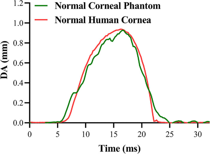
Purpose: To develop a method to fabricate early keratoconus phantom models and evaluate the feasibility of using corneal models for studying the dynamic response of early keratoconus under an air puff.
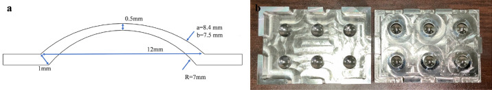
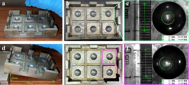
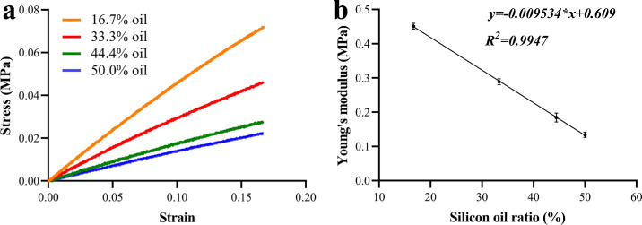
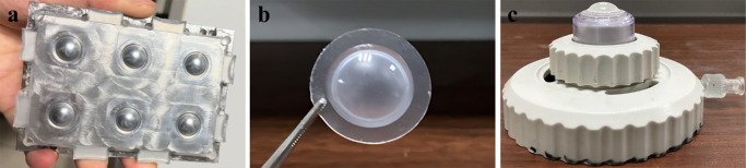
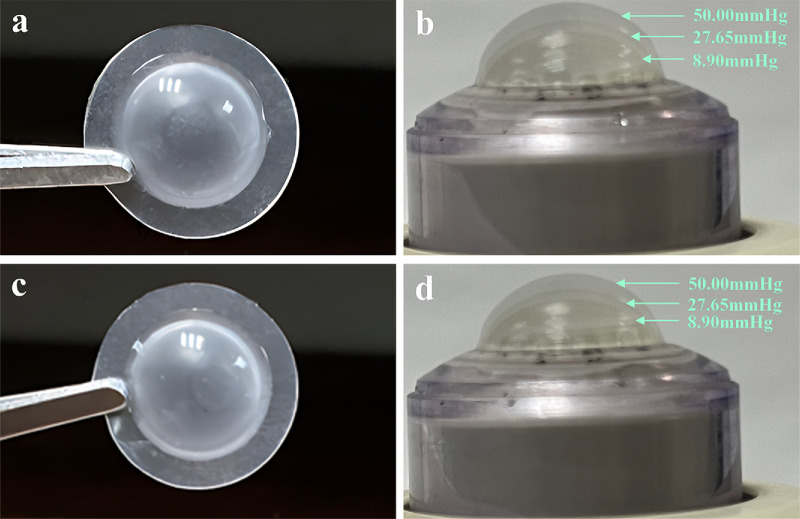
Methods: A corneal mold was designed, and the silicone material was poured into the mold to produce corneal phantoms. Two types of early keratoconus phantoms with reduced mechanical properties in a specific area were prepared using a two-step molding process: the central keratoconus phantom and the paracentral keratoconus phantom. Corneal Visualization Scheimpflug Technology tonometry was performed on the normal corneal phantoms and early keratoconus phantoms, and the corresponding dynamic corneal response (DCR) parameters were recorded.
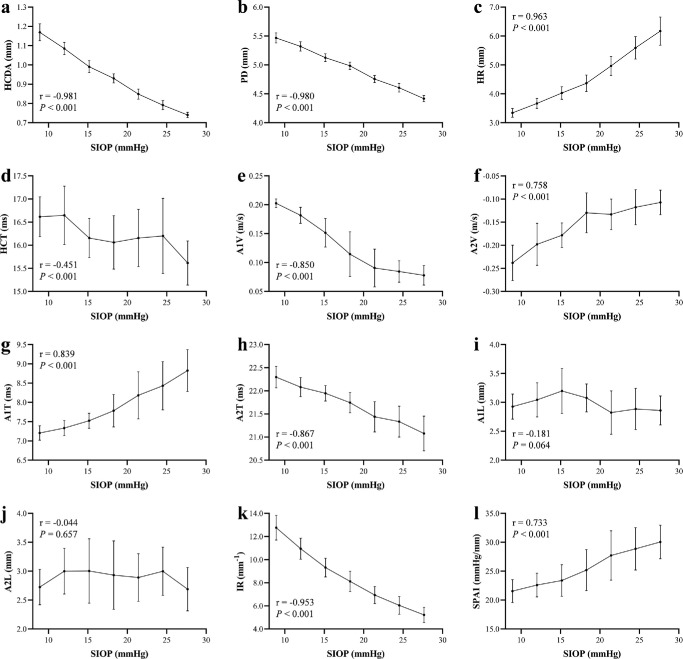
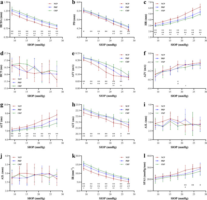
Results: A majority of DCR parameters of the normal corneal phantoms, including deflection amplitude at highest concavity (HCDA), peak distance (PD), radius of curvature (HR), first and second applanation times (A1T and A2T), first and second applanation velocities (A1V and A2V), and the stiffness parameter at the first applanation (SPA1), exhibited trends in response to changes in the simulated intraocular pressure (SIOP) that aligned with experimental results based on ex vivo animal eyes. Significant differences in HCDA, PD, HR, A1V, A2V, A1T, A2T, and integrated radius (IR) were observed between the early keratoconus phantoms and the normal corneal phantoms.
Conclusions: The early keratoconus phantom models fabricated by the present novel approach are feasible for studying the dynamic response of early keratoconus under an air puff.
Translational relevance: This study demonstrated the potential of corneal phantom models for corneal biomechanical studies, which can deepen our understanding of the DCR parameters, and the results will provide valuable information for early diagnosis of keratoconus.
Conflict of interest statement
Disclosure:
Figures
References
-
- Randleman JB, Russell B, Ward MA, Thompson KP, Stulting RD.. Risk factors and prognosis for corneal ectasia after LASIK. Ophthalmology. 2003; 110(2): 267–275. - PubMed
-
- Gomes JAP, Tan D, Rapuano CJ, et al.. Global consensus on keratoconus and ectatic diseases. Cornea. 2015; 34(4): 359–369. - PubMed
-
- Henriquez MA, Hadid M, Izquierdo L.. A systematic review of subclinical keratoconus and forme fruste keratoconus. J Refract Surg. 2020; 36(4): 270–279. - PubMed
-
- Ruberti JW, Sinha Roy A, Roberts CJ. Corneal biomechanics and biomaterials. Annu Rev Biomed Eng. 2011; 13(1): 269–295. - PubMed
-
- Ali NQ, Patel DV, McGhee CNJ.. Biomechanical responses of healthy and keratoconic corneas measured using a noncontact Scheimpflug-based tonometer. Invest Ophthalmol Vis Sci. 2014; 55(6): 3651–3659. - PubMed
MeSH terms
LinkOut - more resources
Full Text Sources
