. 2025 Apr 17;23(4):e3003119.
doi: 10.1371/journal.pbio.3003119. eCollection 2025 Apr.

The length of the G1 phase is an essential determinant of H3K27me3 landscapes across diverse cell types

Affiliations

The length of the G1 phase is an essential determinant of H3K27me3 landscapes across diverse cell types

Abby Trouth et al. PLoS Biol. .

Abstract

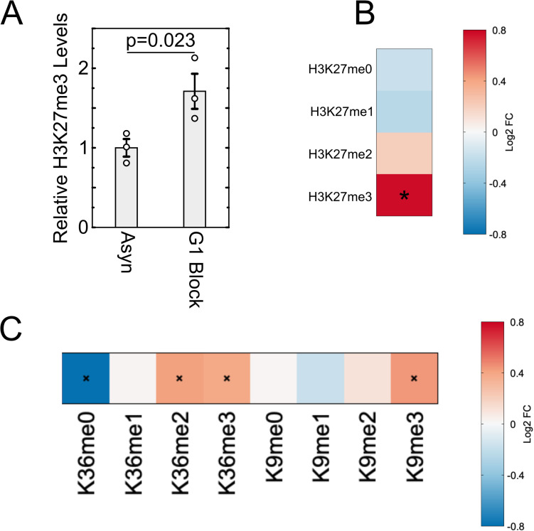
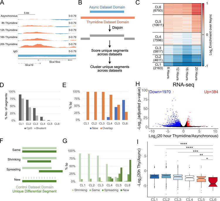
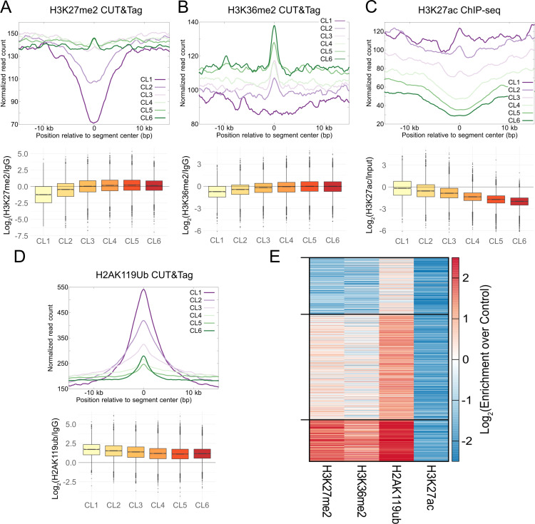
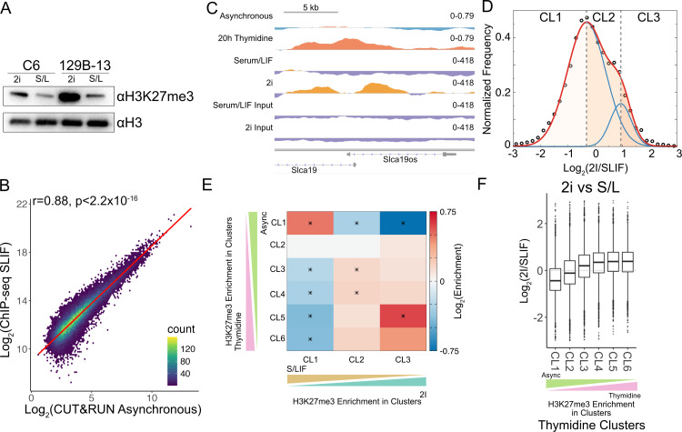
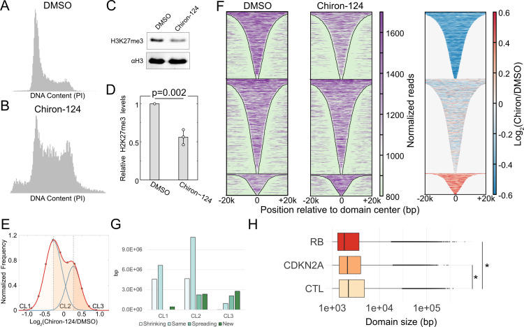
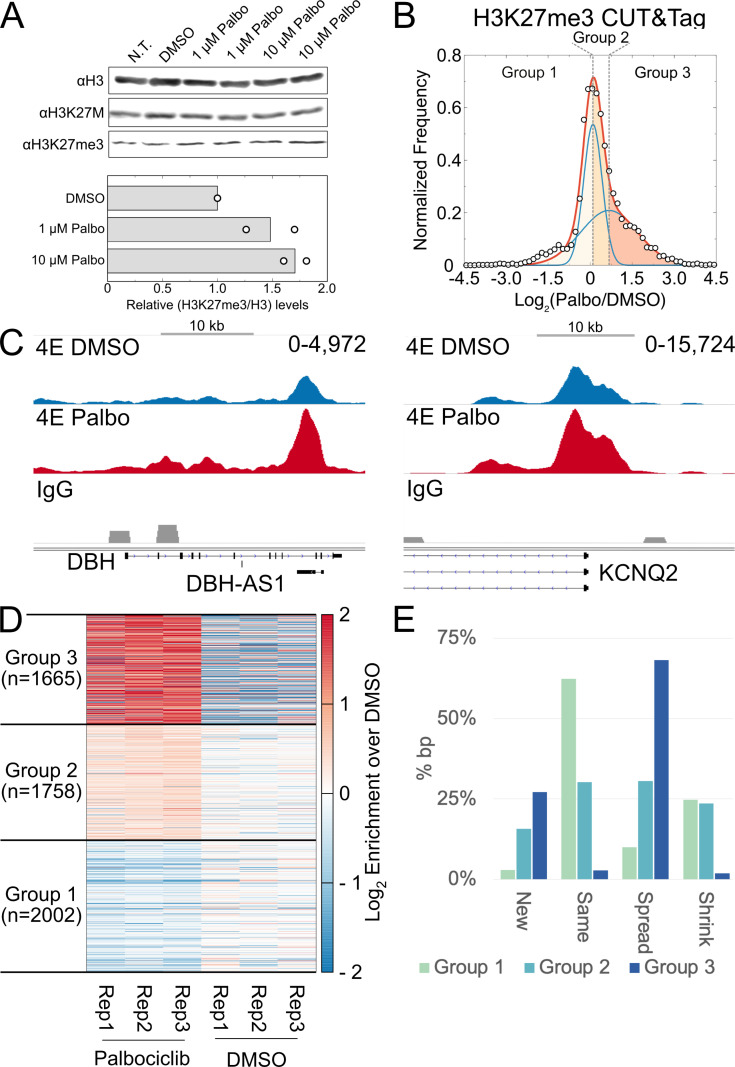
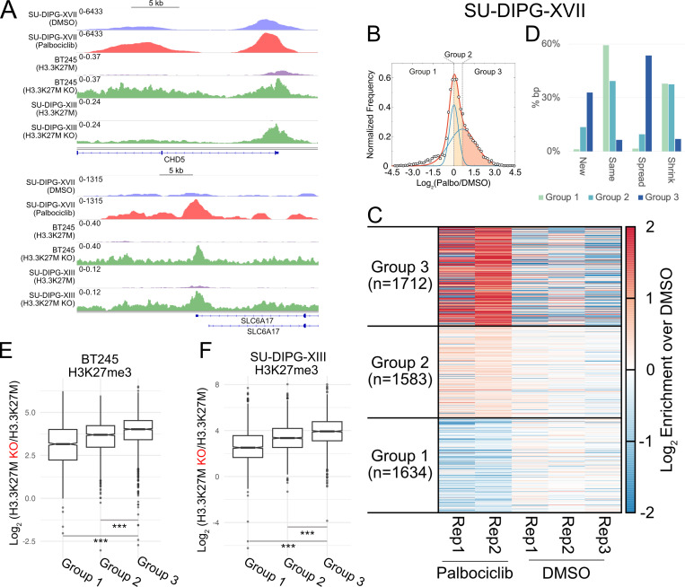
Stem cells have lower facultative heterochromatin as defined by trimethylation of histone H3 lysine 27 (H3K27me3) compared to differentiated cells. However, the mechanisms underlying these differential H3K27me3 levels remain elusive. Because H3K27me3 levels are diluted 2-fold in every round of replication and then restored through the rest of the cell cycle, we reasoned that the cell cycle length could be a key regulator of total H3K27me3 levels. Here, we propose that a short G1 phase restricts H3K27me3 levels in stem cells. To test this model, we determined changes to H3K27me3 levels in mouse embryonic stem cells (mESCs) globally and at specific loci upon G1 phase lengthening - accomplished by thymidine block or growth in the absence of serum (with the "2i medium"). H3K27me3 levels in mESCs increase with G1 arrest when grown in serum and in 2i medium. Additionally, we observed via CUT&RUN and ChIP-seq that regions that gain H3K27me3 in G1 arrest and 2i media overlap, supporting our model of G1 length as a critical regulator of the stem cell epigenome. Furthermore, we demonstrate the inverse effect - that G1 shortening in differentiated human HEK293 cells results in a loss of H3K27me3 levels. Finally, in human tumor cells with extreme H3K27me3 loss, lengthening of the G1 phase leads to H3K27me3 recovery despite the presence of the dominant negative, sub-stoichiometric H3K27M mutation. Our results indicate that G1 length is an essential determinant of H3K27me3 landscapes across diverse cell types.


Conflict of interest statement

The authors have declared that no competing interests exist.

Figures

Fig 1
Fig 1. Global H3K27me3 levels in serum/LIF-grown mESCs increase with G1/S arrest.
(A). Change in relative H3K27me3 levels determined via mass spectrometry across three replicates for cells that underwent double thymidine block with the second block lasting 20 h. (B). Heat map of average fold change across three replicates in H3K27 methylation states in G1 arrested cells versus asynchronous cells. (C). Heat map of fold change in methylation states for H3K36 and H3K9 after a 20-h G1/S arrest for average values across the three replicates. Values for each methylation state were normalized to values obtained from asynchronous cells. Asterisk (*) indicates p < 0.05, p-values calculated using Student’s t test with a one-tailed distribution. The data underlying this figure can be found in S1 Data.
Fig 2
Fig 2. Locus-specific changes in H3K27me3 upon G1/S arrest.
(A). An example of a genomic region showing progressive gain in H3K27me3 upon thymidine treatment. The genomic snapshot was created using IGV, setting the midpoint of the data range as the lower cut-off used in calling domains. Thus, data above midpoint (red) would belong to domains, whereas data below midpoint (blue) would be outside domains. (B). Schematic detailing how non-overlapping segments are defined by comparing H3K27me3 domains for asynchronous and thymidine-treated mESCs. (C). Heatmap of H3K27me3 CUT&RUN enrichment from three replicates combined at four time points of G1/S arrest after performing k-mean clustering with 6 clusters. Each horizontal line of the heatmap represents a unique segment determined by Disjoin. (D). Percentage of the number of segments in each of the 6 clusters containing CpG islands or bivalent promoters. (E). Percentage of bp covered by segments belonging to each of the 6 clusters defined in (C) that overlaps with H3K27me3 domains present in asynchronous mESCs. (F). Schematic detailing four domain changes observed after thymidine arrest. Unique segments from thymidine datasets were classified as “same” when maintaining the same boundaries as an asynchronous domain, “shrinking” when one or more boundaries retract, “spreading” when one or more boundaries extend past those of the asynchronous domain, and “new” for domains not present in asynchronous cells. (G). Percentage of bp covered by segments belonging to each of the four categories of domain changes (shrinking, same, spreading, and new) observed in the 6 clusters defined in (C). (H). Volcano plot where the Log2(Fold Change) in RNA for 20 h Thymidine vs. asynchronous is plotted against -log10(FDR) for genes. Genes with Log2(Fold Change) >1 and FDR < 0.05 are marked in red, whereas genes with Log2(Fold Change) <-1 and FDR < 0.05 are marked in blue. (I). Log2(Fold Change) in RNA of significantly changing genes belonging to each cluster plotted as boxplots. P-values were calculated using Wilcoxon rank sum test and adjusted for multiple testing using Benjamini-Hochberg procedure. The data underlying Fig 2A can be found at accession GSE264214 in GEO, the data underlying Fig 2D, 2E, 2G–I can be found in S2 Data.
Fig 3
Fig 3. H3K27me3 gains due to longer G1 occur at weak nucleation sites.
(A). Enrichment of H3K27me2 CUT&Tag normalized read count averaged over unique segments in each thymidine cluster defined in Fig 2, plotted relative to centers of the unique segments (top). The log2 ratio of the H3K27me2 normalized read count at each segment to the IgG normalized read count is plotted as a boxplot for each thymidine cluster (bottom). (B). Same as (A) for H3K36me2 CUT&Tag. (C) Same as (A) for H3K27ac ChIP-seq. (D). Same as (A) for H2AK119ub CUT&Tag. (E). k-means clustering (k = 3) of log2 enrichment of H3K27me2, H3K36me2, H2AK119ub, and H3K27ac for segments in thymidine clusters 5 and 6 that gain H3K27me3 plotted as a heatmap. H3K27ac is depleted in all clusters, and enrichment of H3K27me2, H3K36me2, and H2AK119ub correlate with each other. The data underlying Fig 3A–E can be found in S3 Data.
Fig 4
Fig 4. Regions with H3K27me3 gain in 2i-grown mESCs overlap with those in G1/S arrest of serum-grown mESC.
(A). Immunoblot for H3K27me3 modification of two mESC cell lines grown in either serum/LIF (S/L or SLIF) media or 2i media. (B). Hexagonal binning of H3K27me3 enrichment at segments belonging to the six clusters defined in Fig 2C from ChIP-seq on the y-axis and CUT&RUN on the x-axis plotted using “geom_hex” in ggplot2. The line of best fit plotted using “geom_smooth” with method “lm” in ggplot2. The Pearson correlation coefficient and the associated p-value is indicated on the top left of the plot. (C). An example of a genomic region (same as shown in Fig 2A) showing gain in H3K27me3 upon thymidine treatment and in 2i compared to Serum/LIF. The genomic snapshot was created using IGV, setting the midpoint of the data range as the lower cut-off used in calling domains. Thus, data above midpoint (red/orange) would belong to domains, whereas data below midpoint (blue/purple) would be outside domains. (D). Distribution of the log2 ratio of H3K27me3 levels in 2i versus serum cells for unique domain segments shown as black circles. The distribution was fitted with the sum of two normal distributions (plotted in red). The individual distributions are plotted in blue. The shaded regions correspond to the three groups based on the log2 ratio: more H3K27me3 in SLIF cells (CL1, leftmost region), equivalent H3K27me3 in SLIF and 2i cells (CL2, center region), and regions with more H3K27me3 in 2i cells (CL3, rightmost region). (E). Extent of overlap of clusters of unique regions from G1/S block of serum grown cells and unique regions defined in (D). Asterisks denote statistical significance determined using hypergeometric test with multiple testing correction (corrected p < 0.05). (F). Log2 ratio of H3K27me3 enrichment of 2i mESC versus serum mESC calculated at unique segments defined in Fig 2. The data underlying Fig 4B, 4D, 4E, and 4F can be found in S4 Data, and data underlying Fig 4C available at accession GSE264216 in GEO.
Fig 5
Fig 5. H3K27me3 domains in HEK293 cells change with accelerated cell cycle timing.
(A). Flow cytometry analysis of DNA content using propidium iodide fluorescence for HEK293 cells that were treated with DMSO for 48 h. (B). Same as (A) HEK293 cells treated with Chiron-124 for 48 h. (C). Immunoblot for H3K27me3 and H3 on acid-extracted histones after 48-h treatment with Chiron-124. (D). Quantification of the modification levels normalized to DMSO-treatment. p-value calculated using two-tailed, homoscedastic student t test. (E). Distribution of the log2 ratio of H3K27me3 levels in Chiron-124 treatment compared to the DMSO treatment for unique domain segments. H3K27me3 levels were determined from the combined reads of two replicates for each condition. Data points are shown as black outlined circles; the sum of the two normal distributions is plotted as red; individual normal distributions are plotted as blue lines. The shaded regions correspond to the three groups based on the log2 ratio: loss of H3K27me3 upon Chiron-124 treatment (CL1, leftmost region), equivalent H3K27me3 in Chiron-124 and DMSO treatment (CL2, center region), and regions with more H3K27me3 in Chiron-124 treatment (CL3, rightmost region). (F). Heatmap of H3K27me3 enrichment at domains defined in DMSO treated cells for DMSO treatment (left) and Chiron-124 treatment (middle), and the log2 ratio of H3K27me3 within domains for Chiron-124 over DMSO (right). (G). Number of bp covered by segments belonging to each of the four categories of domain changes (shrinking, same, spreading, and new) observed in the three clusters shown in the bimodal distribution of H3K27me3 for Chiron-124 versus DMSO cells. (H). Distribution of H3K27me3 domain sizes across ENCODE cell lines with inactivated Rb or CDKN2A deletion or neither. Domain sizes were determined from SEGWAY annotations from ENCODE. *p < 2.2e–16 by Wilcoxon rank sum test. The data underlying Fig 5D–G can be found in S5 Data, with flow cytometry data for Fig 5A and B found in the flow repository public repository at accession FR-FCM-Z96P.
Fig 6
Fig 6. H3K27me3 domains are restored upon G1 arrest in a patient-derived H3.1K27M mutant DMG cell line.
(A). Immunoblot for H3, H3K27M, and H3K27me3 after 72-h treatment with Palbociclib (top). Quantification of the modification levels normalized to DMSO-treatment (bottom). (B). Plot of log2 ratio of H3K27me3 levels in Palbociclib compared to DMSO-treated SU-DIPG-IV cells for unique domain segments. The H3K27me3 levels were determined from combining three replicates for each condition. The distribution was fitted with the sum of two normal distributions (plotted in red). The individual distributions are plotted in blue. The shaded regions correspond to the three groups based on the log2 ratio: loss of H3K27me3 upon treatment with Palbociclib (CL1, leftmost region), equivalent H3K27me3 in Palbociclib and DMSO treatment (CL2, center region), and regions with more H3K27me3 in cells treated with Palbociclib (CL3, rightmost region). (C). Representative H3K27me3 domains from the H3K27me3 gain CL3. (D). Heatmap of H3K27me3 CUT&Tag enrichment at individual replicates for SU-DIPG-IV cells treated with Palbociclib compared to DMSO vehicle control for the 3 clusters defined in (B). (E). Percentage of bp covered by segments belonging to each of the four categories of domain changes (shrinking, same, spreading, and new) observed in the 3 clusters defined in (B). Most domains present in CL1 after Palbociclib treatment maintain the same boundaries as observed in DMSO vehicle control samples. CL3 however, which presents the highest log2 enrichment of H3K27me3 after Palbociclib treatment, is mostly composed of domains with spreading boundaries that extend past those observed in DMSO control samples. The data underlying Fig 6A, 6B, 6D, 6E can be found in S6 Data, and the data underlying Fig 6C available at accession GSE264215 in GEO.
Fig 7
Fig 7. Restored H3K27me3 domains upon G1 arrest in a patient-derived H3.3K27M mutant DMG cell line mirror gains observed upon mutation deletion.
(A). Representative H3K27me3 domains with gains in H3K27me3 upon G1 extension via Palbociclib treatment or knockout of H3.3K27M. (B). Plot of log2 ratio of H3K27me3 levels in Palbociclib versus DMSO-treated SU-DIPG-XVII cells for unique domain segments. H3K27me3 levels were calculated by combining two replicates of Palbociclib treatment and three replicates of DMSO treatment, respectively. Data points are shown as black outlined circles and are fitted with a bimodal distribution shown in red. This distribution is further separated into two normal distributions, plotted in blue. The three shaded regions represent groups with shared changes in H3K27me3: regions that lose H3K27me3 upon Palbociclib treatment (CL1, leftmost region), regions that minimally gain or maintain H3K27me3 upon Palbociclib treatment (CL2, center region), and regions that gain H3K27me3 upon Palbociclib treatment (CL3, rightmost region). (C). Heatmap of H3K27me3 CUT&Tag enrichment for individual replicates of SU-DIPG-XVII cells treated with Palbociclib compared to DMSO vehicle control for clusters defined in (B). (D). Percentage of bp covered by segments belonging to each of the four categories of domain changes (shrinking, same, spreading, and new) observed in the 3 clusters defined in (B). CL1 is composed primarily of maintained H3K27me3 domains between Palbociclib and DMSO treatments, along with some domains that present with shrinking boundaries. CL2 shows a similar trend of same and shrinking domains, with the addition of a small percentage of spreading domains. CL3 in comparison is composed predominantly of spreading domains that push past boundaries present in DMSO-treated cells, pointing to much of the observed H3K27me3 gains in this cluster arising from spreading of the mark. (E and F). Plots of log2 ratio of H3K27me3 levels in WT BT245 cells versus H3.3K27M KO cells (E) and in WT SU-DIPG-XIII cells versus H3.3K27M KO cells (F) for unique domain segments defined from Palbociclib treatment of SU-DIPG-XVII. CL3 shows the most gain of H3K27me3 in both KO BT245 cells and KO SU-DIPG-XIII cells, mirroring the gain that is seen in Palbociclib-treated SU-DIPG-XVII cells. The data underlying Fig 7B–F can be found in S7 Data, with data underlying Fig 7A available at accession GSE264215 in GEO.

Update of

Similar articles

References

    1. Li M, Liu G-H, Izpisua Belmonte JC. Navigating the epigenetic landscape of pluripotent stem cells. Nat Rev Mol Cell Biol. 2012;13(8):524–35. doi: 10.1038/nrm3393 . - DOI - PubMed
    1. Wenger A, Biran A, Alcaraz N, Redó-Riveiro A, Sell AC, Krautz R, et al.. Symmetric inheritance of parental histones governs epigenome maintenance and embryonic stem cell identity. Nat Genet. 2023;55(9):1567–78. doi: 10.1038/s41588-023-01476-x . - DOI - PMC - PubMed
    1. Kim JJ, Kingston RE. Context-specific Polycomb mechanisms in development. Nat Rev Genet. 2022;23(11):680–95. doi: 10.1038/s41576-022-00499-0 . - DOI - PMC - PubMed
    1. Bowman SK, Deaton AM, Domingues H, Wang PI, Sadreyev RI, Kingston RE, et al.. H3K27 modifications define segmental regulatory domains in the Drosophila bithorax complex. Elife. 2014;3:e02833. doi: 10.7554/eLife.02833 . - DOI - PMC - PubMed
    1. Atlasi Y, Stunnenberg HG. The interplay of epigenetic marks during stem cell differentiation and development. Nat Rev Genet. 2017;18(11):643–58. doi: 10.1038/nrg.2017.57 . - DOI - PubMed

LinkOut - more resources