. 2025 Apr 30;147(17):14468-14480.
doi: 10.1021/jacs.5c01276. Epub 2025 Apr 17.

Retinal to Retinal Energy Transfer in a Bistable Microbial Rhodopsin Dimer

Affiliations

Retinal to Retinal Energy Transfer in a Bistable Microbial Rhodopsin Dimer

Ivo H M van Stokkum et al. J Am Chem Soc. .

Abstract

Neorhodopsin (NeoR) is a newly discovered fungal bistable rhodopsin that reversibly photoswitches between UV- and near-IR absorbing states denoted NeoR367 and NeoR690, respectively. NeoR367 represents a deprotonated retinal Schiff base (RSB), while NeoR690 represents a protonated RSB. Cryo-EM studies indicate that NeoR forms homodimers with 29 Å center-to-center distance between the retinal chromophores. UV excitation of NeoR367 takes place to an optically allowed S3 state of 1Bu+ symmetry, which rapidly converts to a low-lying optically forbidden S1 state of 2Ag- symmetry in 39 fs, followed by a multiexponential decay to the ground state on the 1-100 ps time scale. A theoretically predicted nπ* (S2) state does not get populated in any appreciable transient concentration during the excited-state relaxation cascade. We observe an intradimer retinal to retinal excitation energy transfer (EET) process from the NeoR367 S1 state to NeoR690, in competition with photoproduct formation. To quantitatively assess the EET mechanism and rate, we experimentally addressed and modeled the EET process under varying NeoR367-NeoR690 photoequilibrium conditions and determined the EET rate at (200 ps)-1. The NeoR367 S1 state shows a weak stimulated emission band in the near-IR around 700 nm, which may result from mixing with an intramolecular charge-transfer (ICT) state, enhancing the transition dipole moment of the S1-S0 transition and possibly facilitating the EET process. We suggest that EET may bear general relevance to the function of bistable multiwavelength rhodopsin oligomers.


Conflict of interest statement

The authors declare no competing financial interest.

Figures

Figure 1
Figure 1
Absorption spectra showing the progressive photoconversion of the red-absorbing state (NeoR690) into the near UV-absorbing state at 367 nm (NeoR367) by red illumination. Total illumination time for each spectrum as indicated, illumination was at 690 nm at 13 mW/cm2. (Figure adapted from, available under a CC-BY-4.0 license. Copyright 2020 M. Broser et al.).
Scheme 1
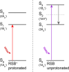
Scheme 1. Energy Level Ordering in Protonated RSB (Left Panel) and Unprotonated RSB (Right Panel)
Figure 2
Figure 2
Evolution-associated difference spectra (EADS) (A) and Decay-associated difference spectra (DADS) (C) estimated with a sequential or parallel analysis of NeoR367 upon excitation at 360 nm. (B,D) Representative traces of data (gray) and fit (black dashed). Note that the time axis is linear until 10 ps, and logarithmic afterward. The estimated lifetimes are 39 fs (black), 0.94 (red), 10 (blue), 93 ps (green), 1.0 ns (magenta) and long-lived (cyan). For presentation purposes the black EADS has been divided by 2. (E) Time-gated spectrum at 100 ps of NeoR690* with direct excitation at 650 nm; (F) FSRS EADS of the NeoR367 S1 state.
Figure 3
Figure 3
Homodimeric arrangement of the NeoR sample as derived from CryoEM. Helices are numbered according to canonical rhodopsins (H1–H7), with the preceding extra helix (H0) found for enzymerhodopsins. Right: representative CryoEM micrograph and selected 2D-class averages.
Figure 4
Figure 4
(A) Transient absorption of NeoR in five conditions monitored at 711 nm upon excitation at 360 nm. Key: experiment 1 (magenta), 2 (green), 3 (cyan), 4 (orange), 5 (gray). Black, red, blue, dark green and purple lines indicate the target analysis fit. (B,C) Dimer fractions (with color key from A) and schematic depiction of the target analysis kinetic schemes for (i) UV–UV dimers; (ii) Red–Red dimers; (iii) UV-Red dimers. Color key: gray S3, blue hot S1, red S1, purple S1′, green P460 photoproduct; black NeoR690* excited state. For case (i), transitions from hot S1, S1 and S1′ to S0 have not been depicted. In case (ii), excitation occurs to a higher excited state NeoR**, which rapidly relaxes to the lowest excited state NeoR*. NeoR** is not further considered in the target analysis. (D) Populations of the states with the color code indicated in (C) that follow from the target analysis under the 5 excitation conditions. Line type key: 1 (dotted), 2 (dot dot dashed), 3 (dot dashed), 4 (dashed), experiment 5 (solid). (E) SADS estimated from the target analysis, with color code identical to panel (D). Note that the SADS of S1 and S1′ are assumed identical and are indicated in red. For presentation purposes the black NeoR690* SADS has been divided by 2, and the S3 SADS has been omitted in panel (E). Both axes in panel (D) are partially linear, and partially logarithmic.

References

    1. Ernst O. P.; Lodowski D. T.; Elstner M.; Hegemann P.; Brown L. S.; Kandori H. Microbial and animal rhodopsins: structures, functions, and molecular mechanisms. Chem. Rev. 2014, 114 (1), 126–163. 10.1021/cr4003769. - DOI - PMC - PubMed
    1. Zhang F.; Vierock J.; Yizhar O.; Fenno L. E.; Tsunoda S.; Kianianmomeni A.; Prigge M.; Berndt A.; Cushman J.; Polle J.; et al. The Microbial Opsin Family of Optogenetic Tools. Cell 2011, 147 (7), 1446–1457. 10.1016/j.cell.2011.12.004. - DOI - PMC - PubMed
    1. Hochbaum D. R.; Zhao Y.; Farhi S. L.; Klapoetke N.; Werley C. A.; Kapoor V.; Zou P.; Kralj J. M.; Maclaurin D.; Smedemark-Margulies N.; et al. All-optical electrophysiology in mammalian neurons using engineered microbial rhodopsins. Nat. Methods 2014, 11 (8), 825–833. 10.1038/nmeth.3000. - DOI - PMC - PubMed
    1. Broser M.; Spreen A.; Konold P. E.; Schiewer E.; Adam S.; Borin V.; Schapiro I.; Seifert R.; Kennis J. T. M.; Bernal Sierra Y. A.; et al. NeoR, a near-infrared absorbing rhodopsin. Nat. Commun. 2020, 11 (1), 5682.10.1038/s41467-020-19375-8. - DOI - PMC - PubMed
    1. Rozenberg A.; Kaczmarczyk I.; Matzov D.; Vierock J.; Nagata T.; Sugiura M.; Katayama K.; Kawasaki Y.; Konno M.; Nagasaka Y.; et al. Rhodopsin-bestrophin fusion proteins from unicellular algae form gigantic pentameric ion channels. Nat. Struct. Mol. Biol. 2022, 29 (6), 592.10.1038/s41594-022-00783-x. - DOI - PubMed

LinkOut - more resources