. 2025 May:115:105707.
doi: 10.1016/j.ebiom.2025.105707. Epub 2025 Apr 16.

An unconventional T cell nexus drives HCK-mediated chronic obstructive pulmonary disease in mice

Affiliations

An unconventional T cell nexus drives HCK-mediated chronic obstructive pulmonary disease in mice

Amy T Hsu et al. EBioMedicine. 2025 May.

Abstract

Background: Chronic obstructive pulmonary disease (COPD) is a heterogeneous inflammatory lung disease leading to progressive, destructive lung function decline, disability and death, and it is refractory to all current treatments. Haematopoietic cell kinase (HCK) is a druggable SRC-family non-receptor protein tyrosine kinase and COPD candidate gene. It is implicated in the chronic and non-resolving inflammation that causes mucosecretory bronchitis and destruction of small airways and alveoli, but how it drives pathophysiology remains obscure.

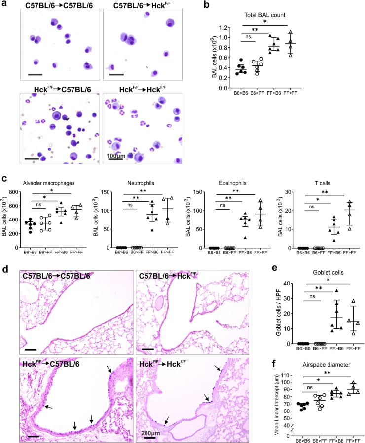
Methods: Studies primarily utilised gene-targeted mice with a gain-of-function mutation in Hck that rendered the enzyme constitutively active. Bone marrow chimeras were established to determine the origin of disease, and the lung disease was investigated using histopathology, morphometry, flow cytometry and single-cell sequencing techniques. Detailed pathways mediating disease pathogenesis were examined using specialised knockout mice.

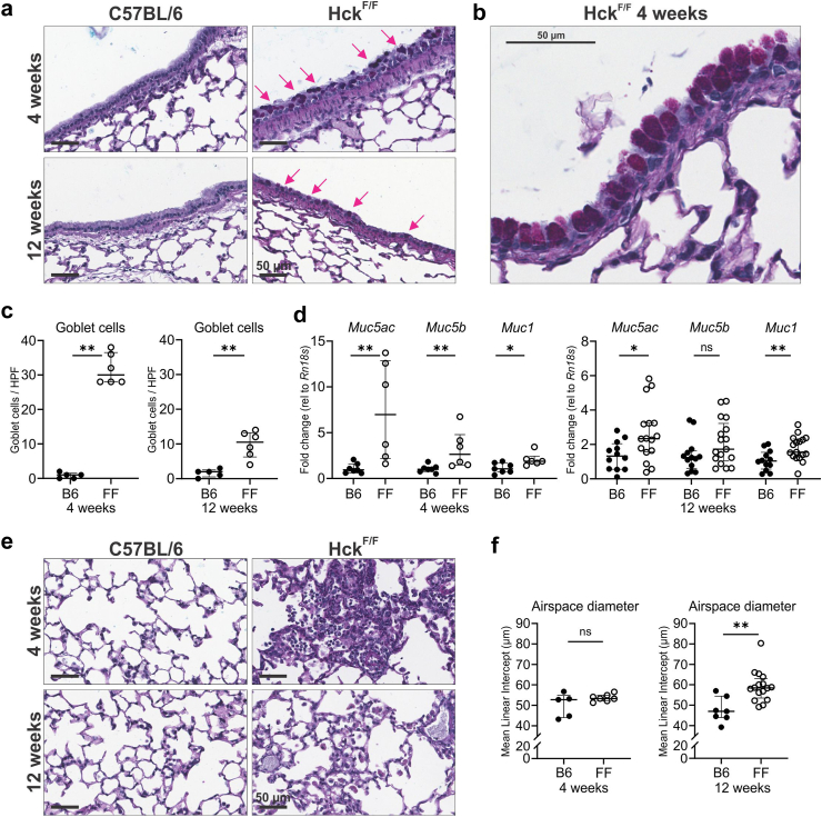
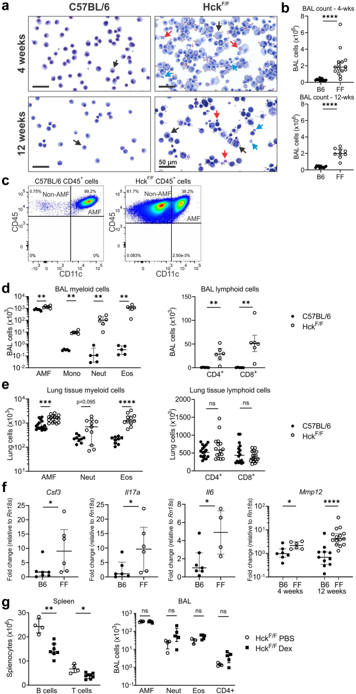
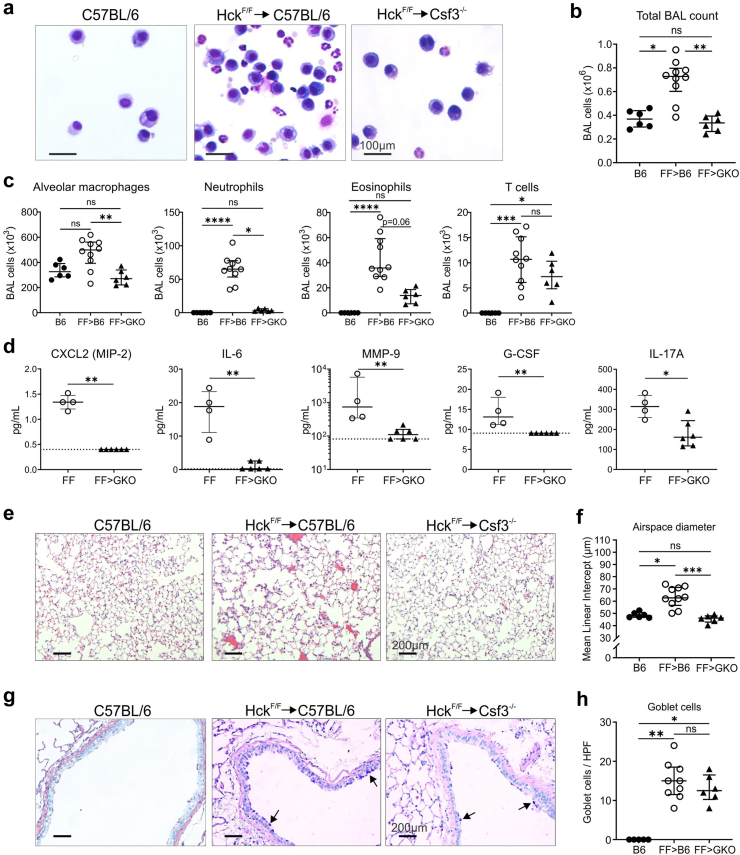
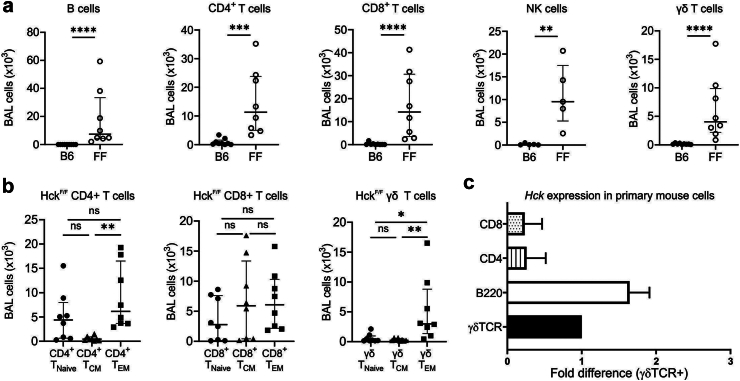
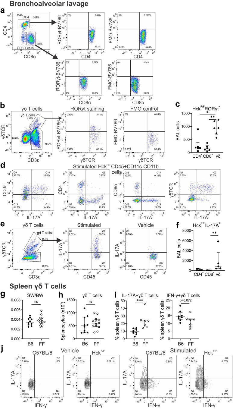
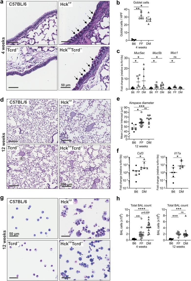
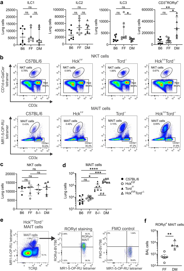
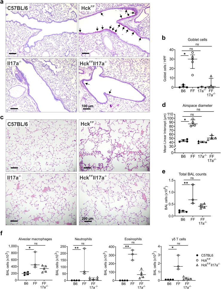
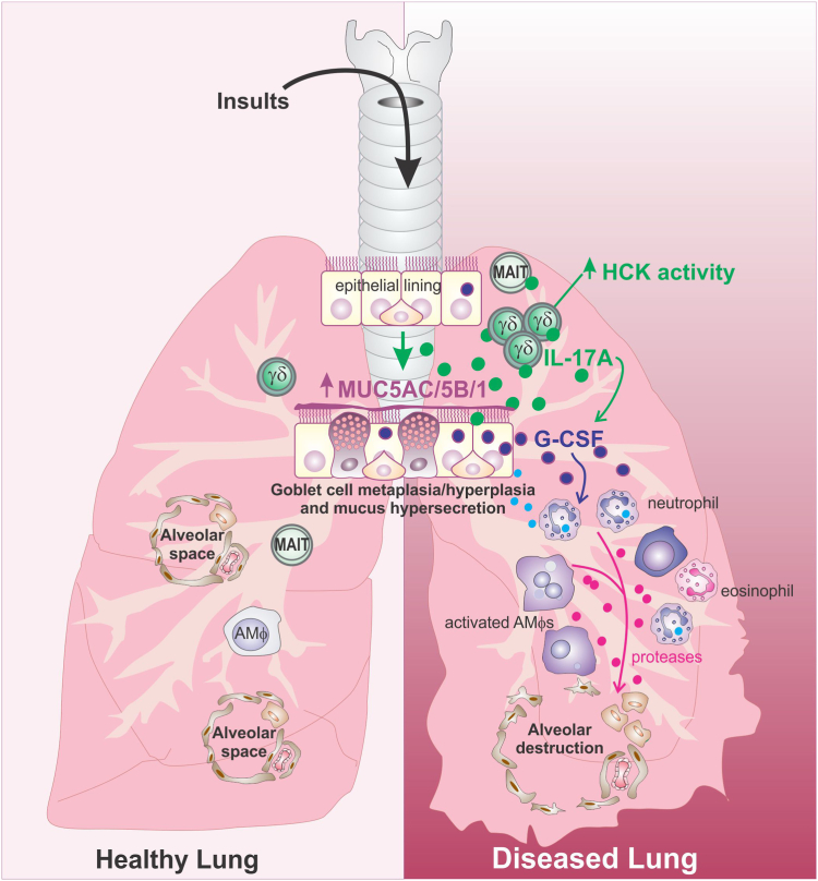
Findings: HckF/F mice developed intense granulocytic mucosecretory inflammation. Bone marrow chimeras revealed that stromal-derived granulocyte-colony-stimulating factor (G-CSF) resulted in lung inflammation and emphysema but not mucus production; while its upstream regulator, interleukin (IL)-17A, itself implicated in emphysema and mucus overproduction, was produced by Vγ6Vδ1 T cells that were recruited to airspaces. Nonetheless, lung disease was unchanged upon genetic deletion of γδ T cells, due to niche-filling expansion of IL-17A-producing mucosal-associated invariant T cells. Strikingly, IL-17A deletion abrogated inflammation, alveolar destruction and mucus overproduction in HckF/F lungs.

Interpretation: These findings highlight the role of HCK as an apical regulator of an unconventional T cell axis that drives IL-17A/G-CSF/granulocyte-mediated pathology in COPD, and underscore the rationale for therapeutically targeting HCK.

Funding: This work received support from the National Health and Medical Research Council Australia, the Victorian Cancer Agency, Melbourne Australia, the Australian Research Council, the Australian Government and the School of Translational Medicine, Monash University, Australia.

Keywords: Chronic obstructive pulmonary disease; Constitutive HCK activation; Granulocytes; IL-17A/G-CSF axis; Mucus-producing goblet cells; Unconventional T cells.


Conflict of interest statement

Declaration of interests ATH and TAG received funding to attend the Australian and New Zealand Society of Immunology conference in 2022 and 2023 respectively; GPA received funding from RAGE Biotechnology, acted as a consultant for RAGE Biotechnology, ENA Respiratory and Pieris Pharmaceuticals, received honoraria from AstraZeneca, GSK and Sanofi, support from Griffith University and Thoracic Society of Australia and New Zealand (TSANZ), participated on Data Safety Monitoring Boards for PACE and INHERIT studies, was a Board Member of TSANZ, and received stock options from ENA Respiratory; and, MLH received grant funding from Lupus Research Alliance and RAGE Biotechnology. The remaining authors declare that they have no competing interests.

Figures

Fig. 1
Fig. 1
HckF/F mice exhibit early-onset increases in goblet cells and lung tissue destruction. (a) Representative images of large airways of AB/PAS-stained lung cross-sections in 4- and 12-wk-old mice depicting mucus-producing goblet cells (arrows) in large airways of HckF/F mice, scale bars represent 50 μm, (b) enlarged image of an independent 4-week-old HckF/F mouse to highlight goblet cell morphology in conducting airway, scale bar represents 50 μm; and, (c) quantitation of goblet cells per high power field (HPF) in large airways, n = 5 B6 and 6 FF mice per group. (d) Lung gene expression analysis of Muc5ac, Muc5b and Muc1 in 4-wk-old mice (n = 7 B6 and 6 FF mice) and 12-wk-old mice (n = 12 B6 and 17 FF mice). (e) Representative images of PAS/AB-stained lung cross-sections from 4- and 12-wk-old C57BL/6 and HckF/F mice, scale bars represent 50 μm; and, (f) quantification of alveolar airspace size by mean linear intercept in 4-wk-old mice (n = 5 B6 and 7 FF mice) and 12-wk-old mice (n = 7 B6 and 16 FF mice). B6 = C57BL/6, FF = HckF/F. Data presented as median ± IQR. ns, not significant, ∗P < 0.05, ∗∗P < 0.01 (Mann–Whitney U test).
Fig. 2
Fig. 2
HckF/F mice display steroid-insensitive lung inflammation. (a) Morphology of BAL cells from 4- and 12-wk-old C57BL/6 and HckF/F mice, scale bars represent 50 μm. Black arrows, alveolar macrophages; red arrows, eosinophils; blue arrows, neutrophils. (b) BAL cell counts of 4-wk-old (n = 13 B6 and 15 FF mice) and 12-wk-old mice (n = 11 B6 and 9 FF mice). (c) Representative flow cytometry pseudocolour plots depicting all viable CD45+ cells in the BAL of 4-wk-old C57BL/6 and HckF/F mice. Flow cytometric quantitation of myeloid and lymphoid cells in (d) BAL (n = 5 B6 and 6 FF mice) and (e) lung tissue (n = 17 B6 and 16 FF mice for alveolar macrophages and lymphoid cells, n = 9 B6 and 11 FF mice for neutrophils and eosinophils) from 4-wk-old mice; AMF, alveolar macrophages; Mono, monocytes; Neut, neutrophils; Eos, eosinophils. (f) Inflammatory gene expression in lung tissue of 4-wk-old mice (n = 7 B6 and 4–6 FF mice); Mmp12 expression in 4-wk-old (n = 7 B6 and 6 FF mice) and 12-wk-old mice (n = 12 B6 and 16 FF mice). (g) HckF/F mice were treated for 4 weeks with PBS (n = 3–4) or 2 mg/kg dexamethasone (Dex) (n = 8) and corticosteroid sensitivity was assessed by analysis of cell populations in spleen (n = 7–8) and BAL (n = 5) by flow cytometry. B6 = C57BL/6, FF = HckF/F. Data presented as median ± IQR. ns, not significant; ∗P < 0.05, ∗∗P < 0.01, ∗∗∗P < 0.001, ∗∗∗∗P < 0.0001 (Mann–Whitney U test).
Fig. 3
Fig. 3
HckF/F haematopoietic cells drive lung inflammation, emphysema and increases in goblet cells. BM chimeras were established as follows: B6 > B6 and B6 > FF denotes C57BL/6 BM transplanted into C57BL/6 or HckF/F mice respectively, FF > B6 and FF > FF denotes HckF/F BM transplanted into C57BL/6 or HckF/F mice respectively. BM chimeras were analysed 16 weeks post-transplantation, n = 6 mice per group except for FF > FF, where n = 4. (a) Morphological analysis of BAL cells, scale bars represent 100 μm; and, (b) total BAL counts of BM chimeras. (c) Flow cytometric quantitation of immune cells in BAL from indicated BM chimeras. (d) Representative AB/PAS-stained lung cross-sections through large airways of the indicated BM chimeras depicting mucus-producing goblet cells (arrows), scale bars represent 200 μm. (e) Quantification of goblet cells numbers in AB/PAS-stained lungs and (f) alveolar diameter by mean linear intercept analysis of H&E-stained lungs. Data presented as median ± IQR. Ns, not significant; ∗P < 0.05, ∗∗P < 0.01 (Kruskal–Wallis test with Dunn's post-test).
Fig. 4
Fig. 4
G-CSF produced by non-haematopoietic cells drives inflammation and emphysema but not goblet cell changes in HckF/F mice. BM chimeras were generated by transplanting HckF/F BM into C57BL/6 or Csf3−/− mice (FF > B6 n = 10 and FF > GKO n = 6 respectively) and were analysed at 12 weeks post-transplant alongside C57BL/6 (B6, n = 6) mice as controls. (a) Morphological analysis of BAL cells, scale bars represent 100 μm; (b) BAL cell counts; and, (c) flow cytometric quantitation of immune cells in BAL. (d) Protein levels in BAL fluid of the FF > GKO chimeras (n = 6) alongside 20-wk-old HckF/F mice (FF, n = 4) by multiplex assay, and IL-17A protein by ELISA. (e) Representative images of H&E-stained sections of the lung parenchyma, scale bars represent 200 μm; and, (f) corresponding quantitation of alveolar airspace size. (g) Representative images of the large airways of AB/PAS-stained lung cross-sections depicting mucus-producing goblet cells (arrows), scale bars represent 200 μm; and, (h) corresponding quantitation of goblet cell numbers. Data presented as median ± IQR. ns, not significant; ∗P < 0.05, ∗∗P < 0.01, ∗∗∗P < 0.001, ∗∗∗∗P < 0.0001 (Kruskal–Wallis test with Dunn's post-test for b, c, f and h, and Mann–Whitney U test for d).
Fig. 5
Fig. 5
γδ T cells are abundant in the BAL of HckF/F mice and have an effector-memory phenotype. (a) Flow cytometric quantification of the indicated lymphoid cells in BAL from 12-wk-old C57BL/6 (B6) and HckF/F (FF) mice, n = 9 B6 and 8 FF mice per group from 2 experiments, except for NK cell analyses, where n = 5 per group from one experiment. (b) Numbers of CD44CD62L+ naïve (Tnaïve), CD44+CD62L+ central memory (TCM) and CD44+CD62L effector memory (TEM) CD4+, CD8+ and γδ T cells in the BAL of HckF/F mice through CD62L vs CD44 gating, n = 8 mice/group from 2 independent experiments. Data presented as median ± IQR, ns = not significant; ∗P < 0.05; ∗∗P < 0.01; ∗∗∗P < 0.001; ∗∗∗∗P < 0.0001 (Mann–Whitney U test for a and Kruskal–Wallis test with Dunn's post-test for b). (c) Gene expression by qPCR of Hck in sorted CD8+ T cells, CD4+ T cells, B220+ B cells and γδTCR + T cells from C57BL/6 mouse spleen. Fold change was calculated relative to Rn18s. Three independent experiments were conducted and the data in each was normalised by expressing the fold difference relative to mouse γδ T cells; pooled data presented as mean ± SEM.
Fig. 6
Fig. 6
γδ T cells in BAL of HckF/F mice are skewed towards IL-17A production. (a) Representative pseudocolour plots of RORγt staining of CD4+ and CD8+ T cells in BAL from HckF/F mice, with FMO control. (b) Representative pseudocolour plots of RORγt staining of γδ T cells in BAL from HckF/F mice, with FMO control. (c) Quantification of RORγt-expressing cells in BAL of 4-wk-old HckF/F mice, n = 6/group. (d) Representative pseudocolour plots depicting IL-17A staining of BAL from HckF/F mice, stimulated for 4 h with PMA/ionomycin. CD45+CD11cCD11b cells were gated and assessed for CD3+, CD4+, CD8+ and γδTCR+ cells. (e) Representative pseudocolour plots depicting IL-17A staining of γδ T cells in BAL from HckF/F mice, stimulated with PMA/ionomycin, or vehicle. (f) Quantification of IL-17A-expressing cells in stimulated BAL from 6-wk-old HckF/F mice, n = 5 mice/group. (g) Spleen weight (SW) as a proportion of body weight (BW) and (h) quantification of γδ T cells in the spleen of 12-wk-old mice by flow cytometry, (n = 10 B6 and 13 FF mice). (i) Proportion of IL-17A+ and IFN-γ+ γδ T cells in spleen after 4 h stimulation with PMA/ionomycin (n = 8 B6 and 7 FF). (j) Representative flow cytometry contour plots of IL-17A and IFN-γ staining of PMA/ionomycin stimulated splenic γδ T cells, with vehicle stimulated splenic γδ T cells shown as control. Data presented as median ± IQR. n = not significant; ∗P < 0.05, ∗∗P < 0.01, ∗∗∗P < 0.001 (Mann–Whitney U test for g–i, and Kruskal–Wallis test with Dunn's post-test for c and f).
Fig. 7
Fig. 7
Deletion of γδ T cells in HckF/F mice does not reduce lung disease or inflammation. (a) Representative images of AB/PAS-stained lung cross-sections from 4-wk-old C57BL/6 (B6), HckF/F (FF), Tcrd−/− and HckF/FTcrd−/− (DM) mice depicting large airways and mucus-producing goblet cells (arrows), scale bars represent 50 μm; and, (b) corresponding quantitation of goblet cells (n = 5 B6, 6 FF and 6 DM). (c) Gene expression of Muc5ac, Muc5b and Muc1 in whole lung tissue of the indicated groups of 4-wk-old mice (n = 7 B6, 6 FF and 6 DM). (d) Representative images of AB/PAS-stained lung cross-sections from the indicated groups of 12-wk-old mice, scale bars represent 200 μm; and, (e) corresponding quantitation of alveolar airspace size (n = 7 B6, 16 FF and 8 DM). (f) Gene expression of Il17a and Csf3 in whole lung tissue of the indicated groups of 4-wk-old mice (n = 7 B6 and 6 DM). (g) Morphological analysis of BAL cells from the indicated 12-wk-old mice; scale bars represent 50 μm. (h) BAL cell counts from the indicated groups of 4-wk-old mice (n = 13 B6, 15 FF and 14 DM) and 12-wk-old mice (n = 11 B6, 9 FF and 13 DM). Data presented as median ± IQR. ns, not significant; ∗P < 0.05, ∗∗P < 0.01, ∗∗∗P < 0.001, ∗∗∗∗P < 0.0001 (Kruskal–Wallis test with Dunn's post-test).
Fig. 8
Fig. 8
Deficiency of γδ T cells in HckF/F mice results in expansion of pulmonary MAIT cells. Quantification of (a) ILC1, ILC2, ILC3, and CD3+RORγt+ cells in digested lung tissue of 12-wk-old C57BL/6 (B6), HckF/F (FF) and HckF/FTcrd−/− (DM) mice by flow cytometry, n = 8/per group. (b) Representative pseudocolour plots of lung tissue cells from the indicated groups of mice stained for NKT and MAIT cells. (c) Flow cytometric quantitation of NKT cells in lung tissue digests of 12-wk-old mice (n = 5 B6, 5 FF, 4 Tcrd−/− (δ−/−) and 6 DM), and (d) flow cytometric quantitation of MAIT cells in lung tissue digests of 12-wk-old mice, n = 8 mice/group. (e) Representative flow cytometry pseudocolour plots of BAL cells from HckF/FTcrd−/− mice gated on MAIT cells and examined for RORγt staining, with FMO control. (f) Flow cytometric quantitation of RORγt + MAIT cells in BAL of HckF/F mice (n = 6) and HckF/FTcrd−/− mice (n = 4). Data presented as median ± IQR. ns, not significant; ∗P < 0.05, ∗∗P < 0.01, ∗∗∗P < 0.001, ∗∗∗∗P < 0.0001 (Kruskal–Wallis test with Dunn's post-test for a, c and d, and Mann–Whitney U test for f). In (d), ##P < 0.01 comparing Tcrd−/− (δ−/−) and HckF/FTcrd−/− mice (Mann–Whitney U test).
Fig. 9
Fig. 9
IL-17A drives lung inflammation, emphysema and goblet cell changes in HckF/F mice. (a) Representative AB/PAS-stained lung cross-sections from 6-wk-old C57BL/6 (B6), HckF/F (FF) and HckF/FIl17a−/− (FF17a−/−) mice depicting mucus-producing goblet cells in large airways (arrows), scale bars represent 100 μm; and, (b) quantification of goblet cells per high power field (HPF) in large airways (n = 4 B6, 5 FF, 3 Il17a−/−, 4 FF17a−/−). (c) Representative H&E-stained lung cross-sections of the indicated groups of 60-wk-old mice; scale bars represent 200 μm; and, (d) corresponding alveolar airspace size; n = 4 mice per group. (e) Total BAL cell counts in indicated groups of 6-wk-old mice, and (f) corresponding flow cytometric quantitation of BAL cell composition (n = 4 B6, 3 FF, 5 FF17a−/−). Data presented as median ± IQR. ns, not significant; ∗P < 0.05, ∗∗P < 0.01 (Kruskal–Wallis with Dunn's post-test).
Fig. 10
Fig. 10
IL-17A-producing unconventional T cells drive HCK-mediated lung disease. γδ T cells and MAIT cells are present in the healthy lung, and together with alveolar macrophages (AMϕ) serve as a first line of defence. In obstructive airways disease, induced by various lung insults, HCK becomes chronically activated, driving an ‘HCK/IL-17 disease endotype’ where HCK-expressing γδ T cells induce goblet cell changes and mucus hypersecretion via IL-17A production, and promote destructive myeloid-rich inflammation via G-CSF.

Similar articles

Cited by

References

    1. Boers E., Barrett M., Su J.G., et al. Global burden of chronic obstructive pulmonary disease through 2050. JAMA Netw Open. 2023;6(12) - PMC - PubMed
    1. Vestbo J., Prescott E., Lange P. Association of chronic mucus hypersecretion with FEV1 decline and chronic obstructive pulmonary disease morbidity. Copenhagen City Heart Study Group. Am J Respir Crit Care Med. 1996;153(5):1530–1535. - PubMed
    1. Takeyama K., Agustí C., Ueki I., Lausier J., Cardell L.O., Nadel J.A. Neutrophil-dependent goblet cell degranulation: role of membrane-bound elastase and adhesion molecules. Am J Physiol. 1998;275(2):L294–L302. - PubMed
    1. Fischer B.M., Voynow J.A. Neutrophil elastase induces MUC5AC gene expression in airway epithelium via a pathway involving reactive oxygen species. Am J Respir Cell Mol Biol. 2002;26(4):447–452. - PubMed
    1. Voynow J.A., Fischer B.M., Malarkey D.E., et al. Neutrophil elastase induces mucus cell metaplasia in mouse lung. Am J Physiol Lung Cell Mol Physiol. 2004;287(6):L1293–L1302. - PubMed

MeSH terms

Substances