. 2025 May 1;112(5):1117-1138.
doi: 10.1016/j.ajhg.2025.03.015. Epub 2025 Apr 16.

Bi-allelic pathogenic variants in TRMT1 disrupt tRNA modification and induce a neurodevelopmental disorder

Stephanie Efthymiou  1 Cailyn P Leo  2 Chenghong Deng  2 Sheng-Jia Lin  3 Reza Maroofian  1 Renee Lin  1 Irem Karagoz  1 Kejia Zhang  2 Rauan Kaiyrzhanov  1 Annarita Scardamaglia  1 Daniel Owrang  4 Valentina Turchetti  1 Friederike Jahnke  4 Kevin Huang  3 Cassidy Petree  3 Anna V Derrick  5 Mark I Rees  6 Javeria Raza Alvi  7 Tipu Sultan  7 Chumei Li  8 Marie-Line Jacquemont  9 Frederic Tran-Mau-Them  10 Maria Valenzuela-Palafoll  11 Rich Sidlow  12 Grace Yoon  13 Michelle M Morrow  14 Deanna Alexis Carere  14 Mary O'Connor  15 Julie Fleischer  15 Erica H Gerkes  16 Chanika Phornphutkul  17 Bertrand Isidor  18 Clotilde Rivier-Ringenbach  19 Christophe Philippe  20 Semra Hiz Kurul  21 Didem Soydemir  22 Bulent Kara  23 Deniz Sunnetci-Akkoyunlu  24 Viktoria Bothe  25 Konrad Platzer  25 Dagmar Wieczorek  26 Margarete Koch-Hogrebe  27 Nils Rahner  28 Ann-Charlotte Thuresson  29 Hans Matsson  29 Carina Frykholm  29 Sevcan Tuğ Bozdoğan  30 Atil Bisgin  31 Nicolas Chatron  32 Gaetan Lesca  32 Sara Cabet  33 Zeynep Tümer  34 Tina D Hjortshøj  35 Gitte Rønde  36 Thorsten Marquardt  37 Janine Reunert  37 Erum Afzal  38 Mina Zamani  39 Reza Azizimalamiri  40 Hamid Galehdari  41 Pardis Nourbakhsh  42 Niloofar Chamanrou  39 Seo-Kyung Chung  43 Mohnish Suri  44 Paul J Benke  45 Maha S Zaki  46 Joseph G Gleeson  47 Daniel G Calame  48 Davut Pehlivan  48 Halil I Yilmaz  49 Alper Gezdirici  49 Aboulfazl Rad  50 Iman Sabri Abumansour  51 Gabriela Oprea  50 Muhammed Burak Bereketoğlu  52 Guillaume Banneau  53 Sophie Julia  53 Jawaher Zeighami  54 Saeed Ashoori  55 Gholamreza Shariati  56 Alireza Sedaghat  57 Alihossein Sabri  56 Mohammad Hamid  58 Sahere Parvas  54 Tajul Arifin Tajudin  59 Uzma Abdullah  60 Shahid Mahmood Baig  61 Wendy K Chung  62 Olga O Glazunova  63 Sigaudy Sabine  63 Huma Arshad Cheema  64 Giovanni Zifarelli  65 Peter Bauer  65 Jai Sidpra  66 Kshitij Mankad  67 Barbara Vona  4 Andrew E Fry  68 Gaurav K Varshney  3 Henry Houlden  69 Dragony Fu  70
Affiliations

Bi-allelic pathogenic variants in TRMT1 disrupt tRNA modification and induce a neurodevelopmental disorder

Stephanie Efthymiou et al. Am J Hum Genet. .

Abstract

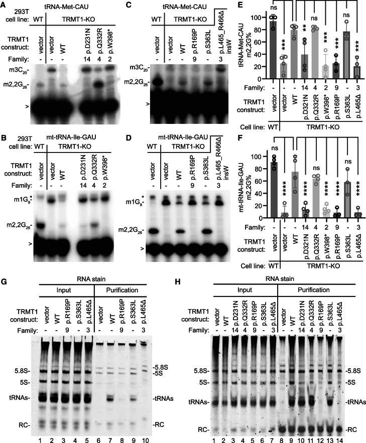
The post-transcriptional modification of tRNAs plays a crucial role in tRNA structure and function. Pathogenic variants in tRNA-modification enzymes have been implicated in a wide range of human neurodevelopmental and neurological disorders. However, the molecular basis for many of these disorders remains unknown. Here, we describe a comprehensive cohort of 43 individuals from 31 unrelated families with bi-allelic variants in tRNA methyltransferase 1 (TRMT1). These individuals present with a neurodevelopmental disorder universally characterized by developmental delay and intellectual disability, accompanied by variable behavioral abnormalities, epilepsy, and facial dysmorphism. The identified variants include ultra-rare TRMT1 variants, comprising missense and predicted loss-of-function variants, which segregate with the observed clinical pathology. Our findings reveal that several variants lead to mis-splicing and a consequent loss of TRMT1 protein accumulation. Moreover, cells derived from individuals harboring TRMT1 variants exhibit a deficiency in tRNA modifications catalyzed by TRMT1. Molecular analysis reveals distinct regions of TRMT1 required for tRNA-modification activity and binding. Notably, depletion of Trmt1 protein in zebrafish is sufficient to induce developmental and behavioral phenotypes along with gene-expression changes associated with disrupted cell cycle, immune response, and neurodegenerative disorders. Altogether, these findings demonstrate that loss of TRMT1-catalyzed tRNA modifications leads to intellectual disability and provides insight into the molecular underpinnings of tRNA-modification deficiency caused by pathogenic TRMT1 variants.

Keywords: TRMT1; disease model; intellectual disability; neurodevelopmental disorder; tRNA modification; zebrafish.


Conflict of interest statement

Declaration of interests M.M.M. and D.A.C. are employees of and may own stock in GeneDx, LLC. R.S. is on the advisory board of Guide Genetics and Egetis Pharmaceuticals.

Figures

Figure 1
Figure 1
Genetic pedigrees of the reported individuals with homozygous TRMT1 variants (A) Pedigrees of the families described. Squares, males; circles, females; black symbols, affected individuals; white symbols, unaffected individuals. Double lines indicate consanguinity. The text below each affected individual describes their alleles with variant alleles in red. (B) Coding exons of the TRMT1 mRNA with variants noted. (C) Schematic indicating the domains of the TRMT1 protein. The red box represents the mitochondrial targeting signal (MTS), while the blue box indicates the class I S-adenosyl-methionine-dependent methyltransferase (SAM MT) domain. The yellow box indicates a C-terminal bipartite nuclear localization signal embedded within a C3H1-type zinc finger (Zn Fn) motif. Variants reported in this study are represented in black, while previously reported variants are in red.
Figure 2
Figure 2
Genetic and phenotypic summary of the reported individuals with homozygous TRMT1 variants (A) Clinical features of the affected individuals with bi-allelic TRMT1 variants. GDD, global developmental delay. (B) Frontal facial photographs of TRMT1 probands showing the most prominent and frequent dysmorphic features of TRMT1-related neurodevelopmental delay. (C) Representative neuroimaging features identified in individuals with intellectual disability. (i) Midsagittal T1-weighted MRI of the brain in a four-year-old boy (F-5) exhibits global (cerebral and cerebellar) atrophy, posterior thinning of the corpus callosum (arrow), and a mega cisterna magna (asterisk). (ii and iii) Midsagittal (ii) and axial (iii) T1-weighted MRI of the brain in a 4-year-old boy (F-8) shows further characteristic features of intellectual disability associated with TRMT1, namely, frontotemporal-predominant cerebral and midbrain atrophy with corresponding ventriculomegaly and uniform thinning of the corpus callosum (not all shown). Note is also made of the right posterior positional plagiocephaly. (iv) Midsagittal T1-weighted brain MRI of 7-year-old boy (F-23) exhibits cerebellar atrophy, a mega cisterna magna (asterisk), and downsloping of the corpus callosum.
Figure 3
Figure 3
TRMT1 variants induce splicing defects and changes in TRMT1 protein levels (A) RT-PCR analysis of RNA from HEK293T cells transfected with TRMT1 minigenes. The presence of additional bands in the WT is attributed to alternative splicing and quantified in Table S7. The splicing schematic is shown for each band to the right. The variant schematic is shown below. Sanger sequencing results showing the correctly spliced WT product with the deleterious variant effect, exon skipping, shown by the presence of only pSPL3 vector. The dotted box represents the short version of exon 5. (B) RT-PCR analysis of RNA from cells transfected with the c.1107−1G>A minigene. The splicing schematic is shown for each band to the right. Exon 10 with 1-bp deletion is represented with a dotted line corresponding to the single-bp deletion. (C) RT-PCR of the c.1194G>A variant. Asterisks represent assay artifacts. The splicing schematic is to the right. Assay design captured part of exon 12 that was correctly spliced in the WT control. (D) Immunoblot of lysates from fibroblast cell lines derived from control (C1) or affected individuals. (E and F) Quantification of TRMT1 levels relative to the control fibroblast cell line after normalization to actin. (G) Immunoblot of lysates from lymphoblast cell lines (LCLs) derived from control WT (C2) or affected individuals. (H) Quantification of TRMT1 levels in LCLs after normalization to actin. n = 3. Error bars represent standard deviation from the mean. Statistical analysis was performed using one-way ANOVA. p ≤ 0.05, ∗∗p ≤ 0.01, ∗∗∗p ≤ 0.001, ∗∗∗∗p < 0.0001; ns, non-significant (p > 0.05).
Figure 4
Figure 4
Human cell lines with bi-allelic TRMT1 variants exhibit a reduction in m2,2G modifications in tRNAs (A and B) Representative gels of primer extension assays to monitor the presence of m2,2G in tRNA-Met-CAU and mt-tRNA-Ile-GAU from the indicated cell lines. m3C20, 3-methylcytosine; m2,2G26, dimethylguanosine; m1G9, 1-methylguanosine. “>” points to labeled oligonucleotide used for primer extension; asterisk denotes background signal. (C–F) Quantification of m2,2G formation by primer extension for the indicated tRNAs. % m2,2G RT block represents the m2,2G stop signal divided by the sum of the m2,2G and read-through m3C or m1G stop signal. The number of replicates is shown in each bar graph with a minimum of two replicates per cell line. Error bars represent standard deviation from the mean. Statistical analysis was performed using one-way ANOVA. For (C) and (D), the mean of each column was compared to the control 1 cell line. For (E) and (F), the mean of each column was compared to the control 2 cell line. p ≤ 0.05, ∗∗p ≤ 0.01, ∗∗∗p ≤ 0.001, ∗∗∗∗p < 0.0001; ns, non-significant (p > 0.05).
Figure 5
Figure 5
TRMT1 protein variants exhibit defects in reconstitution of tRNA-modification activity and interaction with tRNAs (A–D) Representative primer extension gels to monitor the presence of m2,2G in tRNA-Met-CAU and mt-tRNA-Ile-GAU from 293T cell lines transfected with the indicated constructs. m3C20, 3-methylcytosine; m2,2G26, dimethylguanosine; m1G9, 1-methylguanosine. “>” points to oligonucleotide used for primer extension; asterisk denotes background signal. (E and F) Quantification of m2,2G formation by primer extension for the indicated tRNAs. Primer extensions were performed at least three times per variant, and error bars represent the standard error of the mean. Statistical analysis was performed using one-way ANOVA. p ≤ 0.05, ∗∗p ≤ 0.01, ∗∗∗p ≤ 0.001, ∗∗∗∗p < 0.0001; ns, non-significant (p > 0.05). (G and H) Nucleic acid stain of RNAs extracted from the indicated input or purified TRMT1-FLAG samples after denaturing PAGE. The migration pattern of tRNAs, 5.8S, and 5S ribosomal RNA is denoted. The p.L465_R466Δ insW variant is denoted as L465Δ in (E)–(H).
Figure 6
Figure 6
Depletion of Trmt1 in zebrafish causes developmental delay and reduced brain size due to decreased cell proliferation (A) RT-qPCR analysis of trmt1 expression in Cas9-injected control and trmt1 F0 knockout animals at 4 dpf. Expression levels were normalized to 18S and compared to the Cas9 controls. (B and C) LC-MS analysis of m2,2G or m1A in whole larvae or head-only samples. (D) Representative image for Cas9-injected control (Cas9) and trmt1 F0 knockout (F0) larvae at 3 dpf. (E–G) Quantifications of head, eye, and body sizes for Cas9 and trmt1 F0 animals (n = 40 embryos per group). Values are presented as a percentage of the mean value of Cas9 controls. Each dot represents one larva. (H and I) Representative live confocal images of Cas9 and trmt1 F0 larvae in Tg(olig2:dsRed);nacre reporter line at 5 dpf. Images are dorsal view with anterior to the left. dsRed is shown in magenta. Scale bar, 0.1 mm. (J) Quantification of brain regions as depicted in (H) for Cas9 (n = 8 larvae) and F0 (n = 11 larvae) larvae. (K) Schematic illustrating the experimental design: three sgRNAs targeting trmt1 exons were injected into one-cell-stage embryos to generate F0 knockouts. Sexually mature F0 knockouts were bred with WT fish, and the resulting F1 progeny were genotyped to identify inheritable mutant allele carriers. Positive F0 founder carriers were inbred to obtain trans-heterozygous (−/−) F1 knockout progeny. (L) RT-qPCR analysis of trmt1 expression in WT (+/+) control and trmt1 F1 knockout (−/−) larvae at 5 dpf. Experiments were performed with four biological replicates in technical triplicates. Expression levels were normalized to 18S and compared to the WT controls. (M and N) Confocal images of dissected trmt1+/+ (M to M″, n = 5 brains) and trmt1−/− (N to N″, n = 6 brains) larval brain at 5 dpf, stained with anti-acetylated tubulin (Ac-Tub, magenta) and anti-phospho-histone H3 (pHH3, cyan). (M) and (N) show the telencephalon, (M′) and (N′) show the optic tectum and cerebellum, and (M″) and (N″) show the hindbrain. Images are dorsal view with anterior to the left. Brain regions are outlined with a dotted line, and pHH3-positive cells are indicated by white arrowheads. Scale bars, 50 mm. (O) Quantification of the distance between two neuropils as indicated in (M′). (P) Quantifications of areas defined by dotted lines in (M), (M′), and (M″). (Q) Quantification of the number of phospho-histone H3-positive cells in telencephalon and hindbrain. Error bars indicate mean ± SD. For (L), (O), and (P), values are presented as a percentage of the mean value of trmt1+/+ controls. Mean values are displayed at the bottom of each bar. Statistical significance was calculated by unpaired t test with Welch’s correction: ns, non-significant (p > 0.05); p < 0.05, ∗∗p < 0.01, ∗∗∗p < 0.001, ∗∗∗∗p < 0.0001. Tel, telencephalon; Hb, habenula; TeO, optic tectum; Np, tectal neuropil; Cb, cerebellum; Hb, hindbrain.
Figure 7
Figure 7
Transcriptomic analysis of Trmt1-depleted zebrafish larvae reveals differential gene expression related to multiple biological processes and human diseases (A) MA plot highlighting significant DEGs with base-2 log fold-change thresholds of ±1 and a false discovery rate (FDR) of <0.05. Red dots represent upregulated DEGs, while blue dots indicate downregulated DEGs. (B) Heatmap of the 2,000 DEGs, clustered using k-means based on their SD across all samples. Genes were grouped into six clusters, with the number of genes in each cluster shown at top left. (C) Hierarchical tree of the top three enriched GO biological process terms for each cluster, with FDR values displayed before the corresponding GO terms. (D) Hierarchical tree of the top three enriched Alliance Human Disease Ontology (DO) terms for each cluster, with FDR values placed before the DO terms. See Table S9 for detailed lists.

References

    1. Daily D.K., Ardinger H.H., Holmes G.E. Identification and evaluation of mental retardation. Am. Fam. Physician. 2000;61:1059–1070. - PubMed
    1. Knight H.M., Demirbugen Öz M., PerezGrovas-Saltijeral A. Dysregulation of RNA modification systems in clinical populations with neurocognitive disorders. Neural Regen. Res. 2024;19:1256–1261. doi: 10.4103/1673-5374.385858. - DOI - PMC - PubMed
    1. Burgess R.W., Storkebaum E. tRNA Dysregulation in Neurodevelopmental and Neurodegenerative Diseases. Annu. Rev. Cell Dev. Biol. 2023;39:223–252. doi: 10.1146/annurev-cellbio-021623-124009. - DOI - PubMed
    1. Ramos J., Fu D. The emerging impact of tRNA modifications in the brain and nervous system. Biochim. Biophys. Acta. Gene Regul. Mech. 2019;1862:412–428. doi: 10.1016/j.bbagrm.2018.11.007. - DOI - PubMed
    1. Liu J., Strâby K.B. The human tRNA(m(2)(2)G(26))dimethyltransferase: functional expression and characterization of a cloned hTRM1 gene. Nucleic Acids Res. 2000;28:3445–3451. doi: 10.1093/nar/28.18.3445. - DOI - PMC - PubMed

LinkOut - more resources