. 2025 May 8;12(5):ENEURO.0247-24.2025.
doi: 10.1523/ENEURO.0247-24.2025. Print 2025 May.

Characterizing the Diversity of Layer 2/3 Human Neocortical Neurons in Pediatric Epilepsy

Affiliations

Characterizing the Diversity of Layer 2/3 Human Neocortical Neurons in Pediatric Epilepsy

J Keenan Kushner et al. eNeuro. .

Abstract

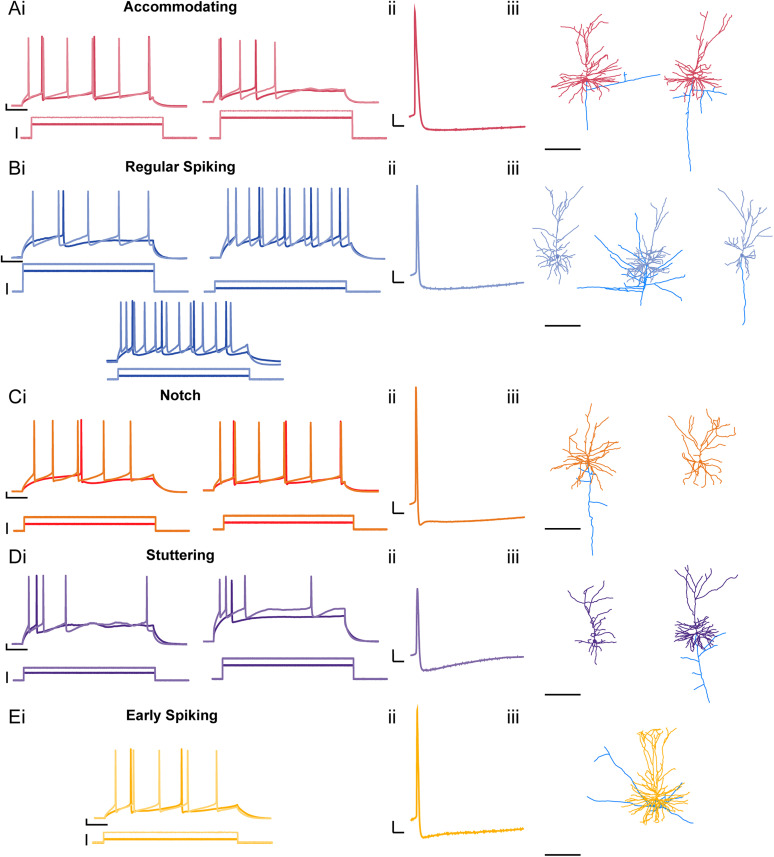
Childhood epilepsy is a common and devastating condition, for which many children still do not have adequate treatment. Some children with drug-resistant epilepsy require surgical excision of epileptogenic brain tissue for seizure control, affording the opportunity to study this tissue ex vivo to interrogate human epileptic neurons for potentially hyperexcitable perturbations in intrinsic electrophysiological properties. In this study, we characterized the diversity of layer L2/3 (L2/3) pyramidal neurons (PNs) in ex vivo brain slices from pediatric patients with epilepsy. We found a remarkable diversity in the firing properties of epileptic L2/3 PNs: five distinct subpopulations were identified. Additionally, we investigated whether the etiology of epilepsy influenced the intrinsic neuronal properties of L2/3 PNs when comparing tissue from patients with epilepsy due to malformations of cortical development (MCDs), other forms of epilepsy (OEs), or with deep-seated tumors. When comparing epileptic with control L2/3 PNs, we observed a decrease in voltage sag and lower maximum firing rates. Moreover, we found that MCD and OE L2/3 PNs were mostly similar indicating that epilepsy etiology may not outweigh the influences of epileptiform activity on L2/3 PN physiology. Lastly, we show that the proconvulsant drug, 4-aminopyridine (4-AP), leads to increased AP half-width, reduced firing rate accommodation, and slower AHPs. These changes imply that 4-AP induces an increase in [K+]o and a resultant increase in AP duration, leading to the release of more excitatory neurotransmitters per action potential, thereby promoting network hyperexcitability.

Keywords: DNET; focal cortical dysplasia; gliosis; intrinsic properties; layer 2/3 pyramidal neurons; tuberous sclerosis.


Conflict of interest statement

The authors declare no competing financial interests.

Figures

Figure 1.
Figure 1.
Putative L2/3 PNs from human epileptic foci have diverse morphology and spiking properties. AiEi, Depolarizing current steps at rheobase (pA; dark trace) and rheobase + 100 pA (2× rheobase, light trace) elicit action potentials (APs) of various shapes and frequencies. Each group was determined based on firing property and AHP shape. AiiEii, Representative trace of each AP from neuron subtype (scale bars: 10 mV, 10 ms). AiiiEiii, Corresponding 3D reconstruction of neuron morphology (x-axis scale bar: 200 µm). Each neuron's location in the representative cortical layers was determined by the respective distance of their soma from pial surface (dendrites: subtype color scheme, axon branches: blue).
Figure 2.
Figure 2.
Putative L2/3 PN subtypes show subtle differences in AP kinetics and firing properties. A, Neuron subtype percentage (%) of the entire population of recorded neurons (ACC, accommodating neurons; RS, regular spiking neurons; Not, notch neurons; ST, stuttering neurons; ES, early spiking neurons). B, Representative traces of L2/3 PN subtype AP (scale bars: 10 mV, 10 ms). C, Overlay of average AP of each neuron subtype. D, Overlay of average phase plot (dV/dT vs voltage) indicating differences in subtype AP kinetics. E, Notch neurons showed the shortest AP half-width with a significant difference compared with accommodating neurons. F, Stutter neurons showed a significant reduction in AP spike amplitude. G, Notch neurons have the shortest AHP latency with a significantly shorter AHP latency compared with RS and ACC neurons. H, FR accommodation ratio of each neuron subtype taken at 2× rheobase indicated ES neurons had a significantly lower chance of accommodating compared with stutter neurons. I, Initial instantaneous frequency ± SEM versus injected current (pA). J, Initial instantaneous frequency was taken at the rheobase + 100 pA current step and indicated significant differences between subtype initial instantaneous firing frequency. K, Final instantaneous frequency ± SEM versus injected current (pA). Scatterplots include mean values ± SD.
Figure 3.
Figure 3.
Epileptic L2/3 PNs with fAHP or mAHP do not differ in their firing rates or firing rate accommodation. A, Representative traces of L2/3 PN AP shapes showing neurons with a (Ai) fAHP followed by mAHP, (Aii) fAHP followed by ADP and mAHP, and (Aiii) mAHP only. B, Overlay of average AP of neuron split by AHP shape. C, Overlay of average phase plots (dV/dT vs voltage) of neuron split by AHP shape. D, There is no significant differences in the maximum depolarization slope of the AP (dV/dT) between subtypes. E, mAHP PNs have a significantly slower repolarization slope (dV/dT) when compared with fAHP PNs. F, AHP latency is shorter for fAHP and ADP PNs as expected compared with mAHP neurons. G, fAHP and ADP PNs show a more positive ΔAHP. H, mAHP PNs have more AP amplitude adaptation compared with ADP PNs. I, mAHP PNs have higher overall voltage sag (%) compared with fAHP PNs. We observe no differences in J, initial instantaneous frequency ± SEM versus injected current (pA); K, mean instantaneous frequency ± SEM versus injected current (pA); L, final instantaneous frequency ± SEM versus injected current (pA); or M, FR accommodation ratio ± SEM versus injected current (pA). N, Percentage (%) of AHP PN based on cell type characterized in Figure 2. f, fAHP; ADP, fAHP-ADP; m mAHP. Scatterplots include mean values ± SD.
Figure 4.
Figure 4.
Epileptic L2/3 PNs show minor differences between etiology, but crucial differences compared with control neurons. A, Representative traces from current (pA) ramp protocol (1,000 pA, 1 s) of L2/3 PNs from control (blue), MCD epileptic (purple), and other epileptic (OE) tissue (green). B, Representative traces from L2/3 PNs from control and epileptic subtype showing the corresponding −250 pA hyperpolarizing step (darker negative trace), rheobase step (darker positive trace), and 2× rheobase (lighter positive trace) depolarizing current step (600 ms steps). C, Percentage (%) of PNs with certain AHP cell type based on control and epilepsy subtype. D, Overlay of average AP of control and epileptic subtype L2/3 PNs. E, Overlay of average phase plots (dV/dT vs voltage) of control and epileptic subtype L2/3 PNs. F, MCD L2/3 PNs have a depolarized AP threshold compared with control. G, OE L2/3 PNs have significantly longer AP half-widths compared with MCD with trends toward longer half-widths in both epileptic subtype PNs compared with control PNs. H, Depolarization slopes (dV/dT) were significantly slower in both epileptic subtypes compared with control. I, AHP magnitude (mV) is significantly larger in both epileptic subtypes compared with control. J, Epileptic L2/3 PNs show a wide variability in input resistance. MCD L2/3 PNs on average have significantly higher input resistances compared with control and OE L2/3 PNs. K, Voltage sag is significantly smaller in both epileptic subtypes compared with control. L, Left, Mean FR ± SEM versus injected current (pA). L, Right, We observed lower max firing rate for both epileptic subtypes compared with control. M, Left, FR accommodation ratio ± SEM versus injected current (pA). M, Right, FR accommodation ratios indicate a lack of FR accommodation by OE L2/3 PNs compared with MCD L2/3 PNs with no differences compared with control. Although, on average, control PNs accommodate their frequency the most compared with the epileptic subtype PNs. Scatterplots include mean values ± SD.
Figure 5.
Figure 5.
4-AP increases L2/3 PN AP half-width and AHP latency and decreases AHP magnitude leading to sustained firing. A, Overlay of a representative AP before and after 4-AP wash on (scale bars: 10 mV, 10 ms). Bi, Overlay of average AP before and after 4-AP wash on. Bii, Overlay of phase plots. C, Input resistance (MΩ) decreased after 4-AP wash on. D, AP half-width increased after 4-AP wash on. E, AP threshold became hyperpolarized after 4-AP wash on. F, AHP magnitude was reduced after 4-AP wash on G, AHP latency got slower after 4-AP wash on. H, AP amplitude was not different after 4-AP wash on. I, Left, Mean FR ± SEM versus injected current (pA). I, Right, No significant difference was found for mean Max FR after 4-AP wash on. J, FR accommodation ratio ± SEM versus injected current (pA) before and after 4-AP wash on at 400, 500, 600, 750, 800, and 850 pA depolarizing current steps indicates a significant absence of accommodation after 4-AP wash on. Scatterplots include mean values ± SD.

References

    1. Aaberg KM, Gunnes N, Bakken IJ, Lund Soraas C, Berntsen A, Magnus P, Lossius MI, Stoltenberg C, Chin R, Suren P (2017) Incidence and prevalence of childhood epilepsy: a nationwide cohort study. Pediatrics 139:e20163908. 10.1542/peds.2016-3908 - DOI - PubMed
    1. Abdijadid S, Mathern GW, Levine MS, Cepeda C (2015) Basic mechanisms of epileptogenesis in pediatric cortical dysplasia. CNS Neurosci Ther 21:92–103. 10.1111/cns.12345 - DOI - PMC - PubMed
    1. Albertson AJ, Yang J, Hablitz JJ (2011) Decreased hyperpolarization-activated currents in layer 5 pyramidal neurons enhances excitability in focal cortical dysplasia. J Neurophysiol 106:2189–2200. 10.1152/jn.00164.2011 - DOI - PMC - PubMed
    1. Andreae LC, Burrone J (2014) The role of neuronal activity and transmitter release on synapse formation. Curr Opin Neurobiol 27:47–52. 10.1016/j.conb.2014.02.008 - DOI - PMC - PubMed
    1. Avoli M, Bernasconi A, Mattia D, Olivier A, Hwa GG (1999) Epileptiform discharges in the human dysplastic neocortex: in vitro physiology and pharmacology. Ann Neurol 46:816–826. 10.1002/1531-8249(199912)46:6<816::AID-ANA3>3.0.CO;2-O - DOI - PubMed