. 2025 Apr 17;16(1):309.
doi: 10.1038/s41419-025-07644-7.

SLC25A42 promotes gastric cancer growth by conferring ferroptosis resistance through enhancing CPT2-mediated fatty acid oxidation

Affiliations

SLC25A42 promotes gastric cancer growth by conferring ferroptosis resistance through enhancing CPT2-mediated fatty acid oxidation

Haoying Wang et al. Cell Death Dis. .

Abstract

Accumulating evidence has shown that the dysfunction of mitochondria, the multifunctional organelles in various cellular processes, is a pivotal event in the development of various diseases, including human cancers. Solute Carrier Family 25 Member 42 (SLC25A42) is a mitochondrial protein governing the transport of coenzyme A (CoA). However, the biological roles of SLC25A42 in human cancers are still unexplored. Here we uncovered that SLC25A42 is upregulated and correlated with a worse prognosis in GC patients. SLC25A42 promotes the proliferation of gastric cancer (GC) cells while suppresses apoptosis in vitro and in vivo. Mechanistically, SLC25A42 promotes the growth and inhibits apoptosis of GC cells by reprograming lipid metabolism. On the one hand, SLC25A42 enhances fatty acid oxidation-mediated mitochondrial respiration to provide energy for cell survival. On the other hand, SLC25A42 decreases the levels of free fatty acids and ROS to inhibit ferroptosis. Moreover, we found that SLC25A42 reprograms lipid metabolism in GC cells by upregulating the acetylation and thus the expression of CPT2. Collectively, our data reveal a critical oncogenic role of SLC25A42 in GCs and suggest that SLC25A42 represent a promising therapeutic target for GC.


Conflict of interest statement

Competing interests: The authors declare no competing interests. Ethics approval and consent to participate: This study has been approved by the Ethics Committee of Tangdu Hospital. Written informed consents have been collected from each GC patients.

Figures
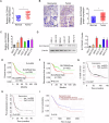
Fig. 1
Fig. 1. SLC25A42 is upregulated and correlates with worse prognosis in patients with gastric cancer.
The detection of SLC25A42 expression was carried out through qPCR (A, n = 40) and IHC (B, n = 385) analysis in paired GC and matched para-cancerous tissues. Scale bar, 20 μm. C, D CSM5 expression was also evaluated in a serial of widely used human GC cell lines (SNU-638, MKN-1, HGC-27, MKN-45, SNU-216) and a normal control gastric cell line (GES-1) through qPCR and Western blot analysis. Kaplan–Meier analysis was performed to compare the survival (E, overall survival; F, disease free survival) of GC patients with low-SLC25A42 and high-SLC25A42 levels. Overall survival (G) and post-progression survival (H) analysis of the gene chip expression database using the online Kaplan-Meier Plotter. I Overall survival analysis of TCGA database using the online UALCAN. *P < 0.05.
Fig. 2
Fig. 2. SLC25A42 promotes the proliferation while suppresses cell death of gastric cancer (GC) cells in vitro.
CCK8 (A) and colony formation (B) elevations for the proliferative potential of gastric cancer (GC) cells with SLC25A42 knockdown or overexpression. C Flow cytometry was utilized to investigate the rates of cell death of GC cells with different SLC25A42 levels (D) The wound healing assay was conducted to compare the migratory abilities of GC cells. E The transwell assay was used to compare the invasive capabilities of GC cells. *P < 0.05; ns, not significant.
Fig. 3
Fig. 3. SLC25A42 promotes in vivo GC tumor growth.
A Subcutaneous tumor models were established using SLC25A42 silencing MKN-1 cells or overexpressing SNU-638 cells and their corresponding control cells (n = 6; Scale bar, 1 cm). The images of excised subcutaneous tumors were shown. B The weights of the tumors were compared in indicated groups. The expression levels of SLC25A42 (C) and Ki-67 (D) in the tissues from subcutaneous nude mice models were assessed by IHC assay. Scale bar, 20 μm. E The occurrence of apoptosis-positive cells in the tissues from subcutaneous nude mice models was assessed by TUNEL assay. Scale bar, 20 μm. F The number of nodules in the lungs from indicated groups was compared by the H&E analysis. Scale bar, 10 μm. *P < 0.05; ns, not significant.
Fig. 4
Fig. 4. SLC25A42 inhibits ferroptosis in GC cells.
A Flow cytometry was utilized to investigate the rates of cell death of GC cells treated with specific inhibitors targeting different types of programmed cell death. 3-Methyladenine (3-MA) targeting autophagy, Z-VAD-FMK (ZVF) targeting apoptosis, Necrostatin-1 (NEC-1) targeting necroptosis. The levels of lipid peroxidation (B) and intracellular Fe2+ (C) were assessed in GC cells exhibiting either SLC25A42 knockdown or overexpression. Scale bar, 10 μm. D IHC staining of 4-hydroxy-2-noneal (4-HNE) in xenograft tumor tissues developed from SLC25A42 knockdown or control MKN-1 cells. F Flow cytometry was utilized to investigate the rates of apoptosis of GC cells. The levels of lipid peroxidation (E) and intracellular Fe2+ (F) were assessed in GC cells treated with the ferroptosis inducer RSL3. Scale bar, 10 μm. *P < 0.05.
Fig. 5
Fig. 5. SLC25A42 enhances fatty acid oxidation (FAO)-supported mitochondrial respiratory activity in GC cells.
Mitochondrial oxygen consumption rate (OCR) (A), membrane potential (B; Scale bar, 5 μm), and intracellular levels of ATP (C) were evaluated in SLC25A42-silencing MKN-1 and SLC25A42-overexpresing SNU-638 cells. Mitochondrial OCR (D), membrane potential (E; Scale bar, 5 μm), and intracellular levels of ATP (F) were evaluated in SLC25A42-silencing MKN-1 and MKN-45 cells treated with inhibitors targeting glucose, fatty acid, and glutamine. G The rate of FAO was assessed using oleic acid (3H-labeled) as the trace in SLC25A42-silencing MKN-1 and SLC25A42-overexpresing SNU-638 cells. *P < 0.05.
Fig. 6
Fig. 6. SLC25A42 promotes GC cell proliferation and inhibit ferroptosis by activation of FAO.
A BODIPY (493/503) staining assay was used to assess lipid amount in SLC25A42-silencing MKN-1 and SLC25A42-overexpresing SNU-638 cells. Scale bar, 20 μm. The amounts of triglycerides (B), phospholipids (C), cholesterol (D), and free fatty acids (E) were determined in GC cells. The levels of NADPH (F) and ROS (G) were determined in GC cells. FI. fluorescence intensity. Scale bar, 20 μm. The CCK8 (H) and colony formation (I) assays for comparing the proliferative potential in SLC25A42-overexpresing SNU-638 cells treated with etomoxir. J Flow cytometry was utilized to investigate the rates of cell death in SLC25A42-overexpresing SNU-638 cells treated with etomoxir. *P < 0.05.
Fig. 7
Fig. 7. SLC25A42 reprograms lipid metabolism in GC cells by upregulating the acetylation and thus the expression of CPT2.
The expressions of key FAO enzymes were detected by qPCR at mRNA level (A) and western blot at protein (B) level. C CPT2 expression was assessed by western blot assay in GC cells treated with CHX. The levels of acyl-CoA (D) and acetylation of CPT2 (E) were assessed by western blot assay in GC cells treated with MG132. F The acetylation of CPT2 was assessed by western blot assay in GC cells treated with SIRT1 inhibitor 3-TYP or activator Compound 5 v. G CPT2 expression was assessed by western blot assay in GC cells treated with SIRT1 inhibitor 3-TYP or activator Compound 5 v. H IHC staining images of CPT2 in GC tissues (left panel) and the correlation between the expressions of SLC25A42 and CPT2 were analyzed (right panel). Scale bar, 20 μm. *P < 0.05; ns, not significant.

References

    1. Smyth EC, Nilsson M, Grabsch HI, van Grieken NC, Lordick F. Gastric cancer. Lancet. 2020;396:635–48. - PubMed
    1. Zeng Y, Jin RU. Molecular pathogenesis, targeted therapies, and future perspectives for gastric cancer. Semin Cancer Biol. 2022;86:566–82. - PubMed
    1. Monzel AS, Enriquez JA, Picard M. Multifaceted mitochondria: moving mitochondrial science beyond function and dysfunction. Nat Metab. 2023;5:546–62. - PMC - PubMed
    1. Liu Y, Sun Y, Guo Y, Shi X, Chen X, Feng W, et al. An overview: the diversified role of mitochondria in cancer metabolism. Int J Biol Sci. 2023;19:897–915. - PMC - PubMed
    1. Genovese I, Carinci M, Modesti L, Aguiari G, Pinton P, Giorgi C. Mitochondria: insights into crucial features to overcome cancer chemoresistance. Int J Mol Sci. 2021;22:4770. - PMC - PubMed

MeSH terms