. 2025 Apr 17;17(1):32.
doi: 10.1038/s41368-025-00360-0.

Dimethyl fumarate modulates M1/M2 macrophage polarization to ameliorate periodontal destruction by increasing TUFM-mediated mitophagy

Affiliations

Dimethyl fumarate modulates M1/M2 macrophage polarization to ameliorate periodontal destruction by increasing TUFM-mediated mitophagy

Liang Chen et al. Int J Oral Sci. .

Abstract

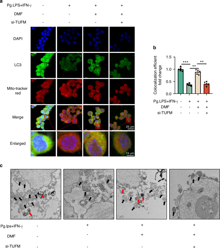
Periodontitis is a common oral disease characterized by progressive alveolar bone resorption and inflammation of the periodontal tissues. Dimethyl fumarate (DMF) has been used in the treatment of various immune-inflammatory diseases due to its excellent anti-inflammatory and antioxidant functions. Here, we investigated for the first time the therapeutic effect of DMF on periodontitis. In vivo studies showed that DMF significantly inhibited periodontal destruction, enhanced mitophagy, and decreased the M1/M2 macrophage ratio. In vitro studies showed that DMF inhibited macrophage polarization toward M1 macrophages and promoted polarization toward M2 macrophages, with improved mitochondrial function, inhibited oxidative stress, and increased mitophagy in RAW 264.7 cells. Furthermore, DMF increased intracellular mitochondrial Tu translation elongation factor (TUFM) levels to maintain mitochondrial homeostasis, promoted mitophagy, and modulated macrophage polarization, whereas TUFM knockdown decreased the protective effect of DMF. Finally, mechanistic studies showed that DMF increased intracellular TUFM levels by protecting TUFM from degradation via the ubiquitin-proteasomal degradation pathway. Our results demonstrate for the first time that DMF protects mitochondrial function and inhibits oxidative stress through TUFM-mediated mitophagy in macrophages, resulting in a shift in the balance of macrophage polarization, thereby attenuating periodontitis. Importantly, this study provides new insights into the prevention of periodontitis.


Conflict of interest statement

Competing interests: The authors declare no competing interests.

Figures

Fig. 1
Fig. 1
DMF ameliorates periodontal tissue destruction. a Macroscopic aspects of the maxilla in mice from control, DMF, ligature, and ligature+DMF groups with volume microscope and 3D reconstruction with Micro-CT. The red lines showed the distance from ACJ to the AC. Representative Micro-CT digitalis images of maxilla alveolar bone surrounding the second upper molar. The square frame showed visual differences in alveolar bone levels between the three groups. b Quantitative analysis of ACJ-AC distance (normalized to the Control group). c Quantitative analysis of bone volume over total volume. d Quantitative analysis of bone mineral density. e, f Representative images of hematoxylin and eosin and tartrate-resistant acid phosphatase stainings of the periodontitis areas. g Quantitative analysis of osteoclast number per view at alveolar bone. Data are presented as mean ± standard error of the mean. *P < 0.05, **P < 0.01, and ***P < 0.001 by one-way analysis of variance followed by Tukey's post hoc test. PDL periodontal ligament
Fig. 2
Fig. 2
DMF modulates M1/M2 macrophage polarization in vivo. a, c Immunofluorescence staining of M1 polarization-related markers (iNOS/CD68) and M2 polarization-related markers (Arginase-1/CD68) and in macrophages across the periodontal region 14 days post-ligature. b, d Semi-quantitative analyses of iNOS+ and Arg1+ ratios in CD68+ cells. Data are presented as mean ± standard error of the mean. *P < 0.05, **P < 0.01, and ***P < 0.001 by one-way analysis of variance followed by Tukey's post hoc test. PDL periodontal ligament
Fig. 3
Fig. 3
DMF alters M1/M2 macrophage polarization in vitro. a IL-1β and tumor necrosis factor-α protein concentration of RAW 264.7 cells tested using enzyme-linked immunosorbent assay. b NO concentration of RAW 264.7 cells tested using Griess assay. c, e Micrographs showing staining of iNOS, DAPI, and Arginase-1 using fluorescence microscopy. (Nucleus: blue, iNOS: red, Argianse-1: green.). d, f Semi-quantitative immunofluorescence analysis for iNOS and Arginase-1 in RAW 264.7 cells. Data are presented as the mean±standard error of the mean and are representative of ≥ 3 independent experiments. *P < 0.05, **P < 0.01, and ***P < 0.001 using T-test and one-way analysis of variance followed by Tukey's post hoc test
Fig. 4
Fig. 4
DMF induces mitophagy in vivo. a Immunofluorescence staining of LC3 and TOM20 in macrophages across the periodontal region 14 days post-ligature. b Co-localization of LC3 and TOM20 was analyzed using ImageJ. Data are shown as the mean±standard error of the mean and are representative of ≥ 3 independent experiments. *P < 0.05, **P < 0.01, and ***P < 0.001 using T-test and one-way analysis of variance followed by Tukey's post hoc test
Fig. 5
Fig. 5
DMF induces mitophagy in vitro. a Micrographs showing staining of LC, DAPI, and Mitotracker using fluorescence microscopy. b Co-localization of LC3 and Mitotracker was analyzed using ImageJ. c Western blot band of Pink1 and P62 expressions in RAW 264.7 cells. d, e Pink1 and P62 levels relative to β-actin. Data are shown as the mean±standard error of the mean and are representative of ≥ 3 independent experiments. *P < 0.05, **P < 0.01, and ***P < 0.001 using T-test and one-way analysis of variance followed by Tukey's post hoc test
Fig. 6
Fig. 6
DMF promotes mitophagy via TUFM in vivo. a Immunofluorescence staining of TUFM and CD68 in macrophages across the periodontal region 14 days post-ligature. b Semi-quantitative analyses of TUFM+ ratio in CD68+ cells. Data are presented as the mean±standard error of the mean and are representative of ≥ 3 independent experiments. *P < 0.05, **P < 0.01, and ***P < 0.001 using T-test and one-way analysis of variance followed by Tukey's post hoc test
Fig. 7
Fig. 7
DMF promotes mitophagy via TUFM in vitro. a Micrographs showing staining of LC, DAPI, and Mitotracker using fluorescence microscopy. b The co-localization of LC3 and Mitotracker was analyzed using ImageJ. c Transmission electron microscopy images of mitochondrial and autophagosome. The red arrows indicate mitochondrion in the characteristic double-membrane autophagosomes, and the black arrows indicate mitochondria: scale bar, 500 nm. Data are presented as the mean±standard error of the mean and are representative of ≥ 3 independent experiments. *P < 0.05, **P < 0.01, and ***P < 0.001 using T-test and one-way analysis of variance followed by Tukey's post hoc test
Fig. 8
Fig. 8
DMF inhibited TUFM degradation via the ubiquitin-proteasome pathway, not through the lysosome pathway. a, d, and e Western blot band of TUFM expression in RAW 264.7 cells. b, f, and g TUFM level relative to β-actin. h Real-time polymerase chain reaction analysis of the gene expression of TUFM. c Ubiquitination experiment of TUFM. Data are presented as the mean±standard error of the mean and are representative of ≥ 3 independent experiments. *P < 0.05, **P < 0.01, and ***P < 0.001 using T-test and one-way analysis of variance followed by Tukey's post hoc test
Fig. 9
Fig. 9
Dimethyl fumarate induces a shift in the polarization phenotype of macrophages in vitro. a IL-1β and TNF-α concentration of RAW 264.7 cells tested using enzyme-linked immunosorbent assay. b NO concentration of RAW 264.7 cells tested using Griess assay. c, e Micrographs showing staining of iNOS, DAPI, and arginase-1 using fluorescence microscopy. (Nucleus: blue, iNOS: red, Argianse-1: green.). d, f Semi-quantitative immunofluorescence analysis for iNOS and Arginase-1 in RAW 264.7 cells. Data are presented as the mean±standard error of the mean and are representative of ≥ 3 independent experiments. *P < 0.05, **P < 0.01, and ***P < 0.001 using T-test and one-way analysis of variance followed by Tukey's post hoc test
Fig. 10
Fig. 10
Schematic diagram for the beneficial effects of dimethyl fumarate against periodontitis through the regulation of TUFM dependent Mitophagy

References

    1. Slots, J. Periodontitis: facts, fallacies and the future. Periodontol 2000.75, 7–23 (2017). - PubMed
    1. Helal, O. et al. Predictors for tooth loss in periodontitis patients: Systematic review and meta-analysis. J. Clin. Periodontol.46, 699–712 (2019). - PubMed
    1. Oh, T. J., Eber, R. & Wang, H. L. Periodontal diseases in the child and adolescent. J. Clin. Periodontol.29, 400–410 (2002). - PubMed
    1. Suvan, J. et al. Subgingival instrumentation for treatment of periodontitis. A systematic review. J. Clin. Periodontol.47, 155–175 (2020). - PubMed
    1. Cekici, A., Kantarci, A., Hasturk, H. & Van Dyke, T. E. Inflammatory and immune pathways in the pathogenesis of periodontal disease. Periodontol. 2000.64, 57–80 (2014). - PMC - PubMed

Publication types

Substances

LinkOut - more resources