. 2025 Apr 17;16(1):3690.
doi: 10.1038/s41467-025-59061-1.

Cryo-EM structures of the small-conductance Ca2+-activated KCa2.2 channel

Affiliations

Cryo-EM structures of the small-conductance Ca2+-activated KCa2.2 channel

Young-Woo Nam et al. Nat Commun. .

Abstract

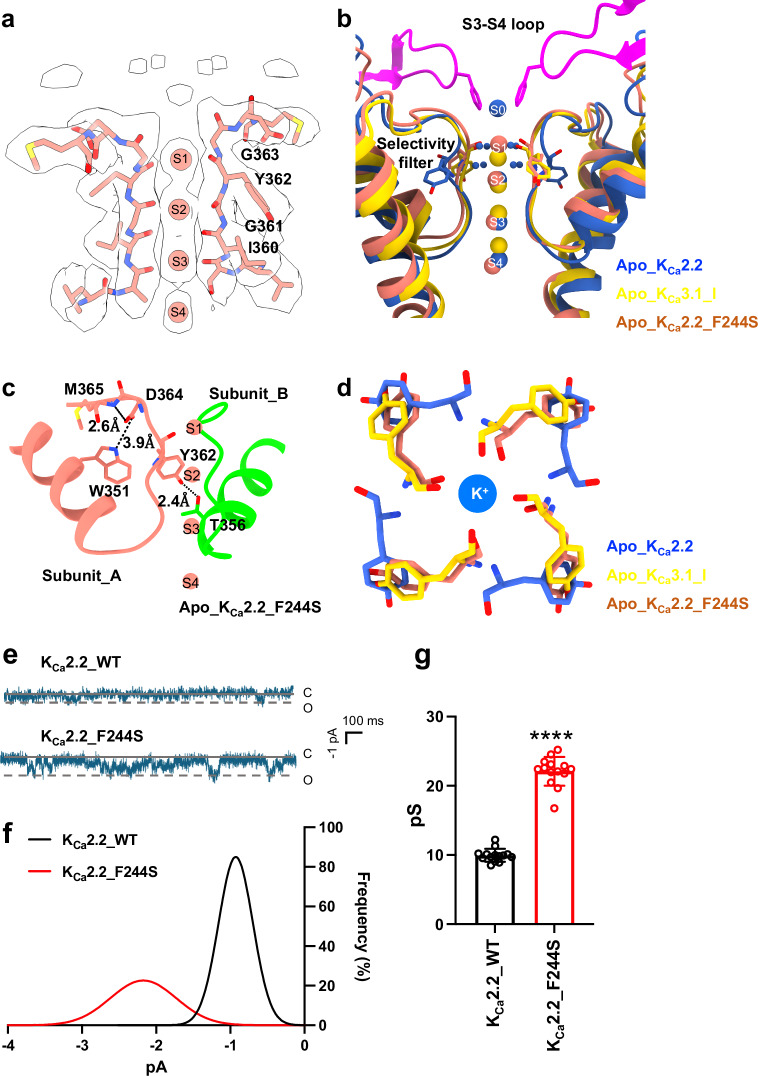
Small-conductance Ca2+-activated K+ (KCa2.1-KCa2.3) channels modulate neuronal and cardiac excitability. We report cryo-electron microscopy structures of the KCa2.2 channel in complex with calmodulin and Ca2+, alone or bound to two small molecule inhibitors, at 3.18, 3.50, 2.99 and 2.97 angstrom resolution, respectively. Extracellular S3-S4 loops in β-hairpin configuration form an outer canopy over the pore with an aromatic box at the canopy's center. Each S3-S4 β-hairpin is tethered to the selectivity filter in the neighboring subunit by inter-subunit hydrogen bonds. This hydrogen bond network flips the aromatic residue (Tyr362) in the filter's GYG signature by 180°, causing the outer selectivity filter to widen and water to enter the filter. Disruption of the tether by a mutation narrows the outer selectivity filter, realigns Tyr362 to the position seen in other K+ channels, and significantly increases unitary conductance. UCL1684, a mimetic of the bee venom peptide apamin, sits atop the canopy and occludes the opening in the aromatic box. AP14145, an analogue of a therapeutic for atrial fibrillation, binds in the central cavity below the selectivity filter and induces closure of the inner gate. These structures provide a basis for understanding the small unitary conductance and pharmacology of KCa2.x channels.


Conflict of interest statement

Competing interests: The authors declare no competing interests.

Figures

Fig. 1
Fig. 1. Cryo-EM structure of apo_KCa2.2 in the Ca2+-bound state.
a Cryo-EM density map with fitted model of apo_KCa2.2 viewed from inside the cell (left), from the plane of the membrane (center), and from the extracellular side (right). Four KCa2.2 subunits are shown in blue, yellow, green and salmon, and CaM is shown in gray. Transmembrane helices S5 and S6 form the ion channel pore, which is surrounded by membrane-embedded helices S1 to S4. b Intracellular view of apo_KCa2.2 (blue/green) superimposed on apo_KCa3.1_1 (yellow/pink, PDB 6cnn) shows channel-CaM interactions that are similar in the two channels. In the cytoplasmic domain, the proximal C-termini of the two channels form HA (Lys402-Thr438 in KCa2.2; Lys293-Thr329 in KCa3.1) and HB (His446-Asn478 in KCa2.2; Ser334-Asn366 in KCa3.1) helices that lie almost parallel to the membrane plane. The HC helices, visible in apo_KCa3.1_1, are invisible in apo_KCa2.2 probably because of flexibility. c Side view of one subunit of apo_KCa2.2 (blue/green) superimposed on a subunit of apo_KCa3.1_1 (yellow/pink). The S3-S4 loop forms a β-hairpin in apo_KCa2.2 (highlighted in magenta), while the S3-S4 loop of apo_KCa3.1_1 is not resolved in PDB 6cnn. d Extracellular view of apo_KCa2.2 (blue) superimposed on apo_KCa3.1_1 (yellow). The four S3-S4 β-hairpins (magenta) form a canopy over the outside of the pore. Phe244 residues (shown as sticks) at the tips of four S3-S4 β-hairpins form an aromatic box with a central opening at the outer end of the pore.
Fig. 2
Fig. 2. Tethering of the S3-S4 β-hairpins to the selectivity filter and 180o flip of the critical tyrosine in the signature sequence in apo_KCa2.2.
a Cryo-EM density map with fitted model of two subunits of apo_KCa2.2 viewed from the plane of the membrane. K+ is seen at positions S0 (at the aromatic box), S2, S3, and S4 within the filter; S1 is missing. Residues in the filter (blue) are highlighted. Extracellular S3-S4 β-hairpins (magenta) from two neighboring subunits are positioned on either side of K+ at S0 in (a, b). Note, the outer filter is widened at the level of Y362-G363. b Each S3–S4 loop is tethered to the filter in the neighboring subunit. Asp364 in the filter of subunit A (blue) forms inter-subunit hydrogen bonds and salt bridge with Arg241, Phe244, and Tyr246 in the S3-S4 loop of neighboring subunit B (green). In addition, an inter-subunit interaction between Trp351 (blue) and Tyr362 (green) in the filter stabilizes the pore. c In apo_KCa3.1_1, Asp255, Val256 and Trp242 (yellow) in the selectivity filter, corresponding to Asp364, Met365 and Trp351 in apo_KCa2.2, form intra-subunit hydrogen bonds. In addition, Tyr253 in apo_KCa3.1_1 (yellow), corresponding to Tyr362 in apo_KCa2.2, forms an inter-subunit hydrogen bond with Thr247 (green). d Extracellular view of the critical tyrosine (Tyr362) in the G[Y/F]G motif of the selectivity filter in apo_KCa2.2 (blue) overlaid on the corresponding aromatic residues in the selectivity filters of the open-conducting conformations of apo_KCa3.1_1 (Tyr253, yellow; PDB 6cnn), apo_KCa3.1_2 (Tyr253, green; PDB 6cno), KVChim (Tyr373, brown, PDB 2r9r), KCa1.1 (Phe279, purple, PDB 5tj6), and AlphaFold_KCa2.2 (Tyr362, gray, https://alphafold.ebi.ac.uk/entry/E9PSQ3). Note, Tyr362 in apo_KCa2.2 is flipped 180° compared to the critical aromatic residues in the other channels.
Fig. 3
Fig. 3. Unique conformation of the selectivity filter in apo_KCa2.2.
a Superimposed outer pores of apo_KCa2.2 (blue-magenta) and apo_KCa3.1_1 (yellow, PDB 6cnn) viewed from the plane of the membrane with two subunits shown. The locations of K+ in the selectivity filters of the two channels are compared. Water molecules are shown as small blue spheres. b Dimensions of the outer pore in apo_KCa2.2 are shown together with the locations of K+. Only two opposite subunits are shown (blue, green). The selectivity filter is widened at the level of Tyr362 and Gly361, above K+ at S2. The S3-S4 β-hairpins of two neighboring subunits (yellow and orange) are shown above the outer end of the selectivity filter. c Pore of apo_KCa2.2 generated by the “Hole” program is shown as a dotted surface. Two opposite subunits are shown. Pore radius: red, <1.15 Å; green, 1.15 to 2.30 Å; blue, >2.30 Å. d Pore radii plotted against distance from the extracellular surface for apo_KCa2.2 (blue), apo_KCa3.1_1 (orange, PDB 6cnn), apo_KCa3.1_2 (green, PDB 6cno), KCa1.1 (purple, PDB 5tj6), and KVChim (brown, PDB 2r9r). Apo_KCa2.2’s outer selectivity filter is wider than the other channels, while its outer vestibule is narrower due to the aromatic (Phe244) box.
Fig. 4
Fig. 4. Untethering of the S3-S4 loop from the selectivity filter and enhanced unitary conductance by the KCa2.2_F244S mutation.
a Cryo-EM density map with fitted model of two opposite subunits of the apo_KCa2.2_F244S mutant. The cryo-EM density corresponding to the S3-S4 β-hairpins seen in apo_KCa2.2 is missing from apo_KCa2.2_F244S. b In the apo_KCa2.2_F244S mutant structure, the S3-S4 β-hairpins and aromatic box are not visible, and the selectivity filter is rearranged into the conformation of a canonical K+ channel selectivity filter. c In the apo_KCa2.2_F244S mutant structure, the selectivity filter is stabilized by intra-subunit hydrogen bonds. d The tyrosine (Y362, salmon) in the G[Y/F]G motif of the selectivity filter in apo_KCa2.2_F244S is flipped 180° relative to apo_KCa2.2 (Y362, blue), and realigns with KCa3.1_1 (Y253, yellow; PDB 6cnn). e Representative traces of single channel recordings of KCa2.2_WT and KCa2.2_F244S at −100 mV. f Representative amplitude histograms of KCa2.2_WT and KCa2.2_F244S. g Summary statistics of single channel conductance values. Data are presented as mean ± SD of single channel conductance values recorded from cells transfected with KCa2.2_WT (n = 14 cells) versus KCa2.2_F244S (n = 14 cells). ****P < 0.0001 compared with KCa2.2_WT (unpaired two-tailed t-test).
Fig. 5
Fig. 5. Interactions of small molecule blocker UCL1684 with KCa2.2.
a Cryo-EM density map refined under C4 symmetry with fitted model of UCL1684_KCa2.2 viewed from the plane of the membrane (left), and from the extracellular side (right). Four KCa2.2 subunits are shown in blue, yellow, green and salmon; CaM is shown in gray; and UCL1684 is shown in magenta. The densities for both the protein and UCL1684 are contoured at σ = 6. b One UCL1684 molecule fitted into its cryo-EM density (magenta) is positioned near the aromatic (Phe244) box at the outer end of the pore. c Representative whole-cell current traces of the KCa2.2_F244S mutant elicited by ramps from −120 mV to 40 mV. d The KCa2.2_F244S mutation abolishes blockade by UCL1684 (500 nM) but retains inhibition by AP14145 (30 μM). e Summary statistics show the KCa2.2_F244S mutant’s responses to UCL1684 and AP14145. Data are shown as mean ± SD of current density values recorded from cells transfected with KCa2.2_F244S (n = 8 cells) in response to the indicated treatments. ****P < 0.0001 compared with control (One-way ANOVA and Tukey’s post hoc tests without adjustments).
Fig. 6
Fig. 6. Interactions of small molecule inhibitor AP14145 with KCa2.2.
a Cryo-EM density map refined under C4 symmetry with fitted model of AP14145_KCa2.2 viewed from the plane of the membrane (left), and from the extracellular side (right). AP14145 density is shown in red. The densities for both the protein and AP14145 are contoured at σ = 6. b Cryo-EM density of AP14145 (red) viewed from the plane of the membrane is shown in the central cavity near Ser359 and Ala384. c Representative whole-cell current traces of the KCa2.2_S359T_A384V mutant elicited by ramps from −120 mV to 40 mV. d The KCa2.2_S359T_A384V double mutations abolish inhibition by AP14145 (30 μM) but retains blockade by UCL1684 (500 nM). e Summary statistics show the KCa2.2_S359T_A384V mutant’s responses to AP14145 and UCL1684. Data are shown as mean ± SD of current density values recorded from cells transfected with KCa2.2_S359T_A384V (n = 6 cells) in response to the indicated treatments. ****P < 0.0001 compared with control (One-way ANOVA and Tukey’s post hoc tests without adjustments).
Fig. 7
Fig. 7. Mechanism of inhibition by AP14145.
a Overlay of apo_KCa2.2 (blue), AP14145_KCa2.2 (orange) and UCL1684_KCa2.2 (green) viewed from the plane of the membrane. AP14145 was docked into the AP14145_KCa2.2 structure guided by the cryo-EM density. The inner gate is defined by Val391 in the transmembrane S6 helices. The inner gate is narrowed in AP14145_KCa2.2 to 6.5 Å compared to 12.3 Å in apo_KCa2.2 and UCL1684_KCa2.2. b In the intracellular view, the inner gate is narrowed in AP14145_KCa2.2 (orange) compared to apo_KCa2.2 (blue) and UCL1684_KCa2.2. c AP14145_KCa2.2’s pore generated by the “Hole” program is shown as a dotted surface with only two opposite subunits shown. Pore radius: red, <1.15 Å; green, 1.15 to 2.30 Å; blue, >2.30 Å. d The pore radii of apo_KCa2.2 (blue), AP14145_KCa2.2 (orange), and UCL1684_KCa2.2 (green) are plotted against distance from the extracellular surface. In this comparison, Phe244 defines the aromatic box formed by the S3-S4 β-hairpins, and Val391 defines the narrowest constriction site of the inner gate. The inner gate is narrowed in AP14145_KCa2.2 compared to the other two structures.

References

    1. Kohler, M. et al. Small-conductance, calcium-activated potassium channels from mammalian brain. Science273, 1709–1714 (1996). - PubMed
    1. Li, W. & Aldrich, R. W. Electrostatic influences of charged inner pore residues on the conductance and gating of small conductance Ca2+ activated K+ channels. Proc. Natl Acad. Sci. USA108, 5946–5953 (2011). - PMC - PubMed
    1. Xia, X. M. et al. Mechanism of calcium gating in small-conductance calcium-activated potassium channels. Nature395, 503–507 (1998). - PubMed
    1. Brown, B. M., Shim, H., Christophersen, P. & Wulff, H. Pharmacology of small- and intermediate-conductance calcium-activated potassium channels. Annu. Rev. Pharmacol. Toxicol.60, 219–240 (2020). - PMC - PubMed
    1. Adelman, J. P., Maylie, J. & Sah, P. Small-conductance Ca2+-activated K+ channels: form and function. Annu. Rev. Physiol.74, 245–269 (2012). - PubMed

MeSH terms