

Upper airway microcirculation remodeling in obstructive sleep apnea is not driven by endothelial activation
- PMID: 40247075
- PMCID: PMC12006345
- DOI: 10.1038/s41598-025-98190-x
Upper airway microcirculation remodeling in obstructive sleep apnea is not driven by endothelial activation
Abstract
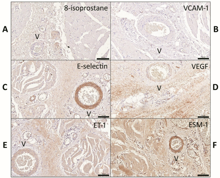
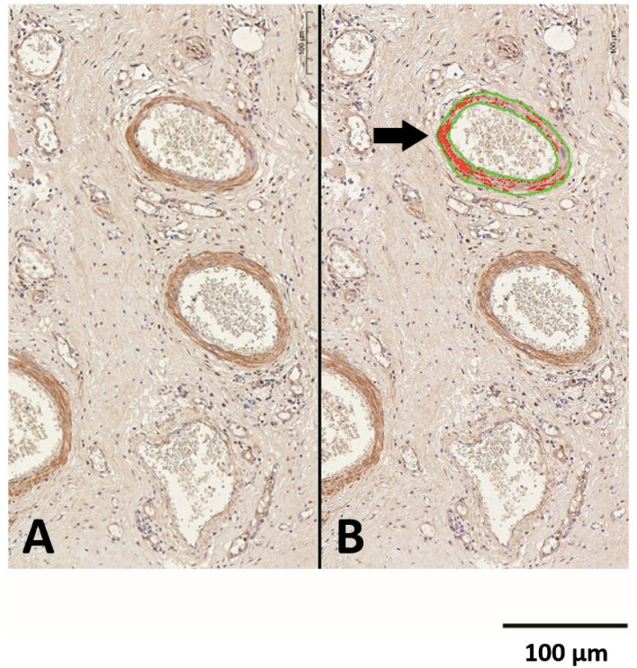
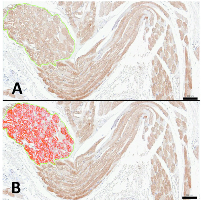
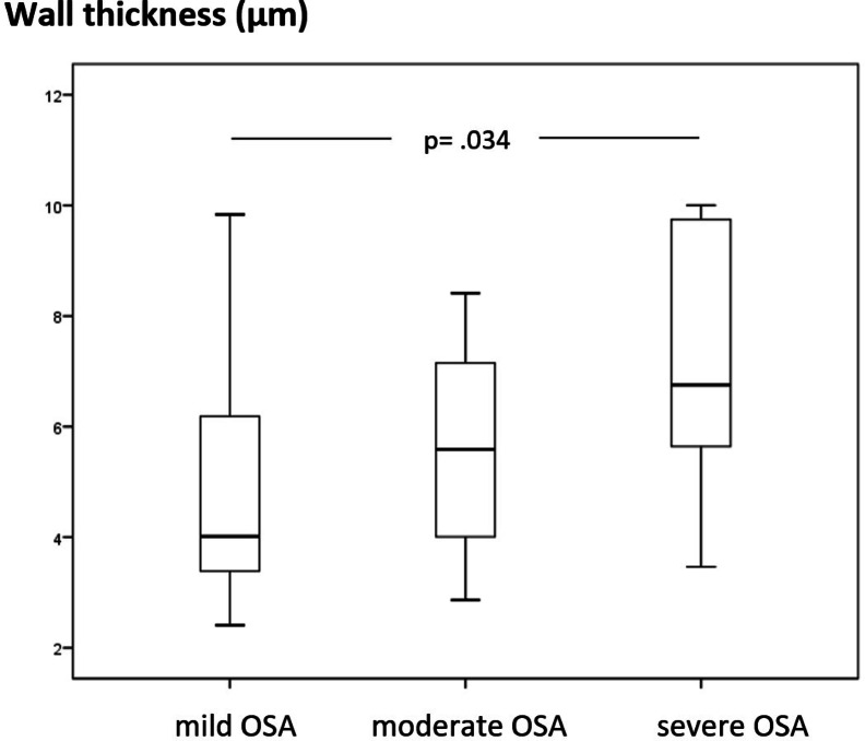
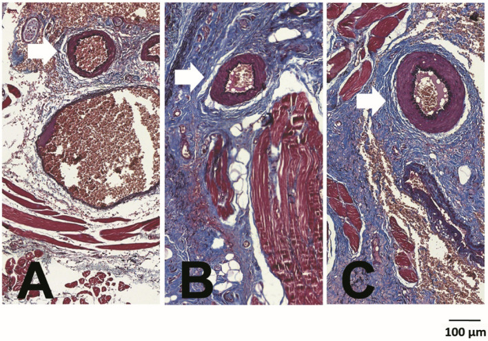
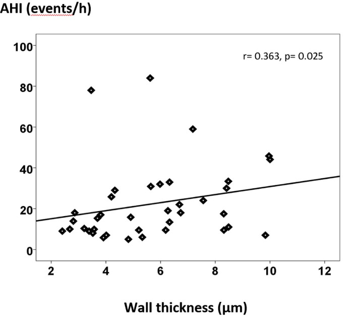
Microcirculation contributes significantly to blood flow resistance, with upper airway microcirculation in obstructive sleep apnea (OSA) affected by endothelial activation, perturbed blood flow shear stress, and snoring-induced tissue vibration. The relevance of these mechanisms on microcirculation response and remodeling remains largely unknown but may influence management decisions in OSA. This study analyzed pharyngeal muscle tissue from non-obese, young adult patients with OSA and chronic heavy snoring. We assessed arteriole morphometry and quantified the expression of endothelial activation markers: 8-isoprostane, vascular cell adhesion molecule-1, E-selectin, vascular endothelial growth factor, endothelin-1, and endothelial cell specific molecule-1. Morphometric analysis of 319 arterioles (mean of 8 vessels per patient) revealed thicker walls in severe OSA compared to mild OSA without lumen reduction, indicating outward hypertrophy, and a positive correlation between the apnea-hypopnea index (a measure of OSA severity) and arteriole wall thickness. However, analysis of 1872 arterioles showed no increase in endothelial activation markers with disease severity, either in the arteriole walls or muscle tissue. This suggests that, in young non-obese adults, severe OSA likely leads to adaptive, mechanically driven microcirculation outward hypertrophy, potentially due to perturbed shear stress, with potential implications for OSA management.
Keywords: Endothelial dysfunction; Microcirculation; Obstructive sleep apnea; Outward hypertrophy; Remodeling; Upper airway.
© 2025. The Author(s).
Conflict of interest statement
Declarations. Competing interests: Michel B. Cahali and Luiz U. Sennes: investors in the Brazilian company Biologix, that markets a home sleep apnea test (not used in this study). Thais Mauad: supported by the Brazilian Research Council (CNPq 304277/2019-3). All the remaining authors declare no conflict of interest.
Figures
References
-
- Somers, V. K. et al. Sleep apnea and cardiovascular disease: An American Heart Association/american College Of Cardiology Foundation Scientific Statement from the American Heart Association Council for High Blood Pressure Research Professional Education Committee, Council on Clinical Cardiology, Stroke Council, and Council On Cardiovascular Nursing. In collaboration with the National Heart, Lung, and Blood Institute National Center on Sleep Disorders Research (National Institutes of Health). Circulation118(10), 1080–1111 (2008). - DOI - PubMed
MeSH terms
Substances
Grants and funding
LinkOut - more resources
Full Text Sources
