. 2025 Jun;10(6):1366-1377.
doi: 10.1038/s41564-025-01998-6. Epub 2025 Apr 17.

Dairy cows develop protective immunity against reinfection with bovine H5N1 influenza virus

Affiliations

Dairy cows develop protective immunity against reinfection with bovine H5N1 influenza virus

Antonio Facciuolo et al. Nat Microbiol. 2025 Jun.

Abstract

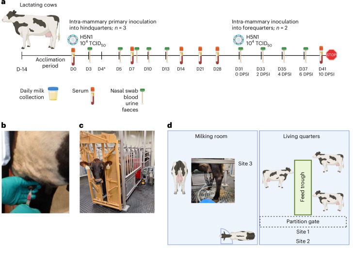
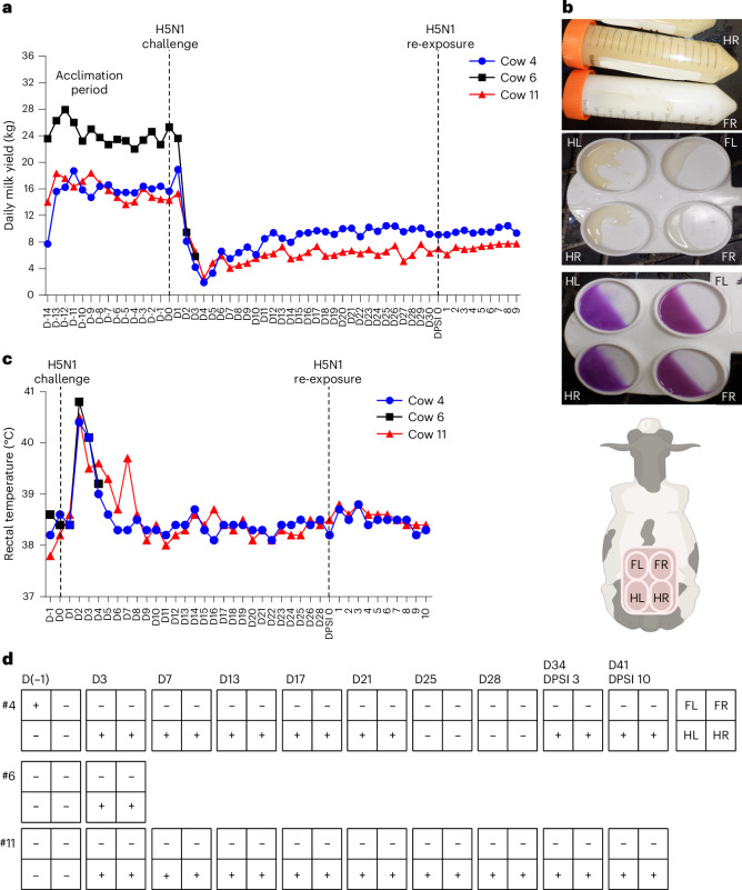
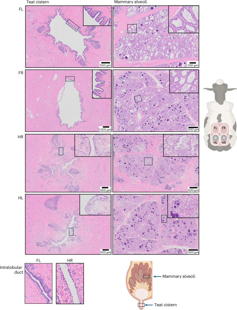
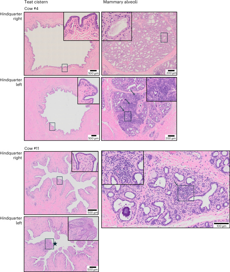
Infection of highly pathogenic avian influenza (HPAI) H5N1 clade 2.3.4.4b in dairy cows causes severe mastitis and milk production losses. Whether cows can develop protective immunity is unclear. Here we infected three lactating cows with HPAI H5N1 genotype B3.13 via the hindquarters of the udder to mimic intra-mammary infection. Inoculated cows displayed clinical responses consistent with affected dairy herds in the United States including virus shedding almost exclusively in inoculated hindquarters that peaked between Days 2-4 post inoculation and gradually declined by Day 21. Histologically, peak virus shedding in milk corresponded with severe acute necrotic mastitis in the inoculated hindquarters but not in the uninoculated forequarters. Two cows were reinfected with HPAI H5N1 virus at unaffected forequarters following resolution of infection. Secondary inoculation did not result in clinical manifestations or virus shedding in milk. Virus-neutralizing antibodies were detected at Day 14 post inoculation in milk with higher titres observed in the inoculated hindquarters relative to the forequarters. We also detected HPAI H5N1 viral RNA in air samples from animal rooms during routine husbandry activity. These data indicate that primary infection via intra-mammary inoculation can generate protective immunity against bovine HPAI H5N1 virus in dairy cows.


Conflict of interest statement

Competing interests: Y.H. is the CEO of PDS (which provided fee-for-service work that included H&E and IHC staining) and is a board-certified pathologist who provided expert interpretation of the histological slides. The other authors declare no competing interests.

Figures

Fig. 1
Fig. 1. Study design.
a, Three late-lactation cows (>300 days in milk) were housed in VIDO Containment Level 3 large animal facility and acclimated for 14 days. At Day 0 (D0) all 3 cows were challenged with 104 TCID50 of bovine H5N1 virus in 1 ml volume inoculated into the teat canal of each hindquarter (b). Blood and nasal secretions, urine and faeces were taken at regular intervals as indicated. Foremilk was collected daily during the morning milking from each of the four quarters. D4 represented peak virus shedding in milk, at which time one cow was euthanized to determine viral tissue distribution and to perform gross and histological examination of tissues. On D31 post primary inoculation, the remaining two cows were inoculated with the same dose into the previously unaffected forequarters. At 10 days post secondary inoculation (DPSI), cows were euthanized and tissues collected for gross and histological examination. c, Dairy cow, during the acclimation period before infection, being prepared for milking using a portable mechanical milker in the dedicated milking room at the VIDO CL3 large animal facility. d, Air samplers were strategically positioned at three distinct sites in the CL3 large animal rooms to simulate farm activities with potential risk of aerosolization and transmission to agricultural workers. Air sampling was conducted during morning milking of the cows which occurred in the milking room (Site 3), during power washing of the living quarters (Site 2), and in the living quarters in the evening (Site 1) to capture bioaerosols generated during these activities. Filter eluent was used to extract RNA and perform RT–qPCR to detect bovine H5N1 viral RNA.
Fig. 2
Fig. 2. Clinical responses following primary inoculation and re-exposure.
a, Cows were milked twice daily. Total milk yield (kg) per day is shown for each cow (#4, 6, 11) during the acclimation period, following bovine H5N1 virus inoculation into the hindquarters, and following re-exposure (DPSI) by inoculation into the forequarters. b, Top: representative milk sample from the hindquarter and forequarter following primary inoculation of bovine H5N1 virus into the hindquarters. Hindquarter milk appeared thickened and yellow in contrast to the normal consistency and colour of milk collected from the forequarters. Middle: representative milk sample collected from each quarter illustrating differences in colour and consistency of hindquarter milk and forequarter milk. Bottom: representative image of the CMT performed on the samples shown in the middle panel. c, Daily rectal temperatures from each cow. d, CMT results shown for select timepoints in the study. The four squares represent each individual quarter of a cow (forequarters, top row; hindquarters, bottom row). FL, forequarter left; FR, forequarter right; HL, hindquarter left; HR, hindquarter right. Source data
Fig. 3
Fig. 3. Histological examination of the teat cistern and mammary alveoli following primary inoculation.
Cow #6 was euthanized at 4 days post inoculation. Representative H&E staining is shown for the teat cistern and mammary alveoli for each quarter, including an intralobular duct from the forequarter and hindquarter. Inoculated hindquarters (HR, HL) show severe acute necrotic mastitis with alveoli filled with proteinaceous material mixed with fat droplets. Images were captured at ×200 magnification, insets are digital magnifications of the boxed area. Cow schematic illustrating anatomical location of each quarter of the udder; mammary gland schematic shows tissue sampled for histological examination. Select figure elements were generated using BioRender.com.
Fig. 4
Fig. 4. Immunohistochemical staining for influenza A following primary inoculation and virus detection in milk.
a, Cow #6 was euthanized at 4 days post inoculation. Representative IHC staining (dark brown colour) for influenza A in the teat cistern and mammary alveoli. Cytoplasmic staining for influenza A indicated by black arrows. Sloughed cells in the mammary alveoli of the left hindquarter showing positive staining. Images were captured at ×200 magnification, insets are digital magnifications of the boxed area. b,d, Virus detection in milk by RT–qPCR. Each sample was tested in duplicate, with the mean Ct value converted to TCID50 equivalent per ml (represented by each data point) by interpolating from a standard curve generated by downtitrating bovine H5N1 virus in milk. c,e, Infectious virus titres in milk. Each data point represents the TCID50 value calculated using the Spearman and Karber algorithm. Cow schematic generated using BioRender.com. Source data
Fig. 5
Fig. 5. Histological examination of the teat cistern and mammary gland following re-exposure.
Cow #4 and #11 were euthanized at 10 days post secondary inoculation (Day 41 post primary inoculation). Representative H&E staining of the teat cistern is shown for the hindquarters of both cows. Representative H&E images of the mammary alveoli from the hindquarters are pictured, showing multifocal lymphoplasmacytic to histiocytic inflammation in the stroma (black arrows). Cow #11 teat cistern shows squamous metaplasia of the epithelium (black star). Images were captured at ×200 magnification, insets are digital magnifications of the boxed area.
Fig. 6
Fig. 6. Antibody responses.
a,c,f, NP-reactive antibodies in serum (a) and milk stripped from individual quarters (c,f) were measured using the IDEXX Influenza A Ab test; S/P ratios < 0.6 were deemed positive. Each data point represents the calculated S/P ratio. Virus-neutralizing (VN) antibody titres in serum (b) and milk stripped from individual quarters (d,g) were quantified using the microneutralization assay. Each data point represents the highest dilution of serum or milk that protected cells from CPE in at least 2 out of 4 replicates. Haemagglutinin inhibition (HAI) antibody titres were measured using the haemagglutination assay for milk stripped from individual quarters (e,h). Each data point represents the reciprocal of the last dilution of milk with complete haemagglutination inhibition, performed in duplicate. Source data
Extended Data Fig. 1
Extended Data Fig. 1. Gross pathology of mammary gland from Cow #6.
Cow #6 was euthanized at 4 days post-inoculation which coincided with peak virus shedding in milk. The mammary tissue of the left hindquarter appeared grossly mildly hyperemic when compared to the other quarters (marked by asterisk). Milk in the forequarters had a grossly normal appearance (black arrow) relative to milk in the hindquarters was thick and discolored (white arrow).
Extended Data Fig. 2
Extended Data Fig. 2. Histological examination of the teat cistern and mammary gland following re-exposure.
Cows #4 and #11 were euthanized at 10 days post secondary inoculation (Day 41 post primary inoculation). Representative H&E staining of teat cistern and mammary alveoli for the forequarters. Images were captured at 200X magnification, insets are digital magnifications of the boxed area.

References

    1. Kandeil, A. et al. Rapid evolution of A(H5N1) influenza viruses after intercontinental spread to North America. Nat. Commun.14, 3082 (2023). - PMC - PubMed
    1. Ramey, A. M. et al. Highly pathogenic avian influenza is an emerging disease threat to wild birds in North America. J. Wildl. Manage. 86, e22171 (2022).
    1. Giacinti, J. A. et al. Avian influenza viruses in wild birds in Canada following incursions of highly pathogenic H5N1 virus from Eurasia in 2021–2022. mBio15, e0320323 (2024). - PMC - PubMed
    1. Peacock, T. et al. The global H5N1 influenza panzootic in mammals. Nature637, 304–313 (2025). - PubMed
    1. Ulloa, M. et al. Mass mortality event in South American sea lions (Otaria flavescens) correlated to highly pathogenic avian influenza (HPAI) H5N1 outbreak in Chile. Vet. Q.43, 1–10 (2023). - PMC - PubMed

MeSH terms

LinkOut - more resources