Review
. 2025 Jun;22(6):1167-1190.
doi: 10.1038/s41592-025-02647-w. Epub 2025 Apr 17.

Imaging 3D cell cultures with optical microscopy

Affiliations
Review

Imaging 3D cell cultures with optical microscopy

Huai-Ching Hsieh et al. Nat Methods. 2025 Jun.

Abstract

Three-dimensional (3D) cell cultures have gained popularity in recent years due to their ability to represent complex tissues or organs more faithfully than conventional two-dimensional (2D) cell culture. This article reviews the application of both 2D and 3D microscopy approaches for monitoring and studying 3D cell cultures. We first summarize the most popular optical microscopy methods that have been used with 3D cell cultures. We then discuss the general advantages and disadvantages of various microscopy techniques for several broad categories of investigation involving 3D cell cultures. Finally, we provide perspectives on key areas of technical need in which there are clear opportunities for innovation. Our goal is to guide microscope engineers and biomedical end users toward optimal imaging methods for specific investigational scenarios and to identify use cases in which additional innovations in high-resolution imaging could be helpful.


Conflict of interest statement

Competing interests: J.T.C.L. is a cofounder, equity holder and board member of Alpenglow Biosciences. A.K.G. is a cofounder and equity holder of Alpenglow Biosciences. N.L.A. and Y.W. are cofounders and equity holders of Altis Biosystems. B.S.F. is an inventor on patents and patent applications related to human organoid differentiation and disease modeling (US20200377860A1, WO2019222559A1). B.S.F. holds ownership interest in Plurexa. The remaining authors report no conflicts of interest.

Figures

Fig. 1.
Fig. 1.. Illustration of 3D cell culture models with 2D and 3D microscopy.
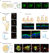
In this article, four types of 3D cell culture are discussed: advanced 2D cultures, spheroids and organoids, organ-on-chip systems, and slice cultures. Two-dimensional microscopy provides a projection view (path-averaged), while 3D microscopy offers “optically sectioned” images, enabling accurate characterization of volumetric morphologies, including intricate internal structures. Example images of colon culture models are shown at the bottom: advanced 2D crypts with central stem cells slightly protruding above the surface (a projection of the 3D images captured by confocal fluorescence microscopy) , colonic organoids (imaged by epifluorescence microscopy) , colon-on-a-chip (detached crypts settled horizontally on a glass slide and imaged with epifluorescence microscopy) , and colon slice cultures (10-μm thin section imaged with epifluorescence microscopy) . (green: stem/proliferative cells; red: differentiated cells; blue: nuclei) (inspired by BioRender.com)
Fig. 2.
Fig. 2.. Major challenges for microscopy of 3D cell cultures.
To illustrate some of the basic processes that make optical microscopy challenging in 3D cell cultures, a beam of light is shown being focused to a localized spot beneath a sample surface (i.e. on the order of 200-microns deep). Living cells and tissues consist of diverse components and interfaces, such as lipid membranes, organelles, cytoplasm, cell filaments, and fluids that all exhibit differences in refractive index. These heterogeneous refractive-index distributions lead to light scattering and aberrations (i.e., changes in photon propagation paths) as light transits through the tissue. These challenges are greater in 3D cell cultures compared to 2D cultures, as 3D cultures are typically larger and have more structural complexity. Light scattering is a pseudo-random process in which light is dispersed in various directions after interacting with small refractive objects. The behavior of any single photon is difficult to predict due the stochastic nature of light scattering. The accumulation of multiple scattering events over millions of photons leads to a reduction in signal at the focus and an increase in a “blur” of undesired background light that reduces image contrast (here defined as signal to background ratio, or SBR). For relatively thin and/or transparent specimens in which this scattering can be limited to an acceptable degree, refractive-index heterogeneities can still result in significant wavefront aberrations. Wavefront aberrations, arising from the refraction of light as it passes through irregular interfaces with different refractive indices, cause the shape of the focus to be distorted and enlarged, which degrades the image quality. In the absence of such aberrations, the size and shape of the beam focus should be close to the ideal limit (i.e., the “diffraction limit”) as predicted by optical diffraction theory. As light propagates within tissue, light is absorbed exponentially as a function of depth. However, at microscopic length scales (< 1 mm deep), the effects of scattering and aberrations typically dominate over the effects of absorption. Other challenges with imaging 3D cultures include the need for uniform staining of the sample, avoiding aberrations and scattering from the sample holder/substrate (if the optical setup requires the light path to transmit through the substrate), minimizing phototoxicity and photobleaching, maintaining ideal environmental conditions for live-culture growth and imaging, and the need for a long working distance to image deeply within larger specimens, especially when using high numerical aperture (NA) objectives that typically have shorter working distances. Note that NA is related to the total range of angles at which light is focused or collected, where a higher NA enables higher spatial resolution. Higher objective magnification is often used as a proxy for higher resolution; however, it is important to note that NA and magnification are distinct parameters and that NA is ultimately the driver of spatial resolution.
Fig. 3.
Fig. 3.. Optical diagram, general characteristics and examples of common 2D and 3D microscopy methods used to image 3D cell culture models.
The performance of six classes of microscopy techniques is roughly generalized and compared for six parameters: optical-sectioning ability (volumetric ability), phototoxicity and photodamage, speed, cost, ease-of-use, and spatial resolution. Example images: transmission light microscopy: intestinal organoid ; epifluorescence microscopy: ileum organoid ; confocal fluorescence and multiphoton microscopy: colonic organoids ; light-sheet fluorescence microscopy: brain organoid
Fig. 4.
Fig. 4.. Imaging 3D cell cultures used in diverse research domains
(a) Developmental biology. Spatial lineage analysis is performed in cerebral organoids using time-lapse imaging with LSFM over several days. Nuclei positions and division events are monitored to elucidate the morphogenesis process. (Scale bar: 100 μm) (b) Infection Biology. The infection behavior of bacteria in intestinal organoids is observed using LSFM, helping to uncover the mechanism of bacterial translocation. (c) Pharmacology. The efficacy of a drug to mitigate radiation-therapy damage is assessed by measuring villus heights in a gut-on-a-chip model. Confocal microscopy results are shown with and without pre-treatment. (Scale bar: 100 μm) (d) Cancer Biology. The killing effects of engineered T cells is tracked in patient-derived tumor organoids using confocal microscopy. (Scale bar: 30 μm; time: hr: min) (inspired by BioRender.com)
Fig. 5.
Fig. 5.. Principles of aberration correction and scattering mitigation techniques for 3D cell cultures.
(a) Generic illustration of AO microscopy, demonstrating aberration detection using either a wavefront sensor or sensorless methods, and correction via a deformable mirror. The example images are of organoids captured by AO-based LSFM before and after aberration correction (green: dynamin; magenta: clathrin). (b) The principle of two-sided illumination/collection in an LSFM system that uses four different combinations of objectives to image different parts of the sample and to avoid the effects of scattering/aberrations at deeper tissue regions. The final image can be generated through a computational image fusion algorithm. (inspired by BioRender.com)
Fig. 6.
Fig. 6.. Principles and applications of multi-scale imaging in 3D cell cultures
(a) An example workflow using multi-scale imaging in which low-resolution imaging is first used to rapidly screen an array of 3D cultures to identify regions of interest, followed by high-resolution imaging of specific structures/regions for detailed quantitative analysis. (b) In one example, a custom-designed multi-well plate, which incorporates micro-mirrors, is used with a conventional microscope equipped with switchable objectives. Initially, simple widefield TLM (2D imaging) is used to record the position of each organoid, followed by the application of moderate-resolution LSFM to detect rare cell-cluster events. High-resolution LSFM is then used to carefully characterize those rare structures (scale bar: 30 μm) (c) In another example, a multi-scale “hybrid” OTLS microscope enables whole cleared mouse brains to be rapidly screened within several hours using a low-resolution imaging path to identify the locations of brain metastases, followed by detailed high-resolution interrogation of small regions of interest containing those metastases for quantitative analysis. (scale bar: 1 mm for low-resolution imaging and 100 μm for high-resolution imaging) . (inspired by BioRender.com)
Fig. 7.
Fig. 7.. Principles of highly multiplexed imaging and their application in 3D cell cultures.
(a) An example workflow for cyclic staining and imaging of fixed samples typically involves staining 3 - 4 protein targets with antibodies, followed by photobleaching and re-staining after each imaging round to enable the detection of multiple targets across cycles. Bottom part is an example multiplexed image of a thin retinal organoid section with 32 “tissue units” in different colors . (b) For live samples, hardware techniques such as hyperspectral imaging can be applied to capture multiple labels. The mixed signals are computationally unmixed to create a final multiplexed image. (c) For virtual staining, AI models are used to predict the staining of multiplexed targets from label-free (or low-plex) images

References

    1. Kapałczyńska M et al. 2D and 3D cell cultures - a comparison of different types of cancer cell cultures. Archives of Medical Science 14, 910–919 (2018). - PMC - PubMed
    2. This review provides a comparison between 2D and 3D cell cultures, as well as among different types of 3D cell cultures.

    1. Warmflash A, Sorre B, Etoc F, Siggia ED & Brivanlou AH A method to recapitulate early embryonic spatial patterning in human embryonic stem cells. Nature Methods 11, 847–854 (2014). - PMC - PubMed
    1. Wang Y et al. Self-renewing Monolayer of Primary Colonic or Rectal Epithelial Cells. Cell Mol Gastroenterol Hepatol 4, 165–182.e167 (2017). - PMC - PubMed
    1. Wang Y, Sims CE & Allbritton NL Human 2D Crypt Model for Assaying Intestinal Stem Cell Proliferation and Differentiation. Analytical Chemistry 94, 9345–9354 (2022). - PMC - PubMed
    1. van der Putten C et al. Protein Micropatterning in 2.5D: An Approach to Investigate Cellular Responses in Multi-Cue Environments. ACS Applied Materials & Interfaces 13, 25589–25598 (2021). - PMC - PubMed

MeSH terms

LinkOut - more resources