. 2025 Apr 17;31(1):139.
doi: 10.1186/s10020-025-01198-2.

Diprovocim protects against the radiation-induced damage via the TLR2 signaling pathway

Affiliations

Diprovocim protects against the radiation-induced damage via the TLR2 signaling pathway

Duo Fang et al. Mol Med. .

Abstract

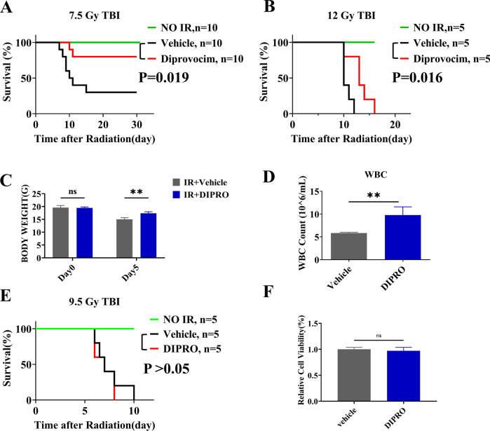
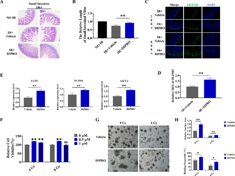
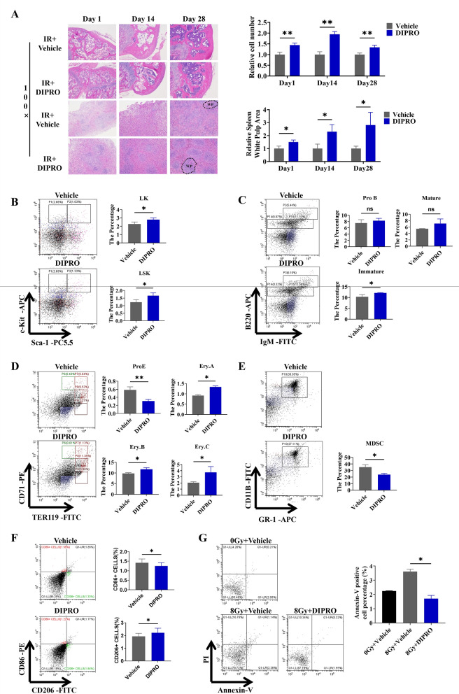
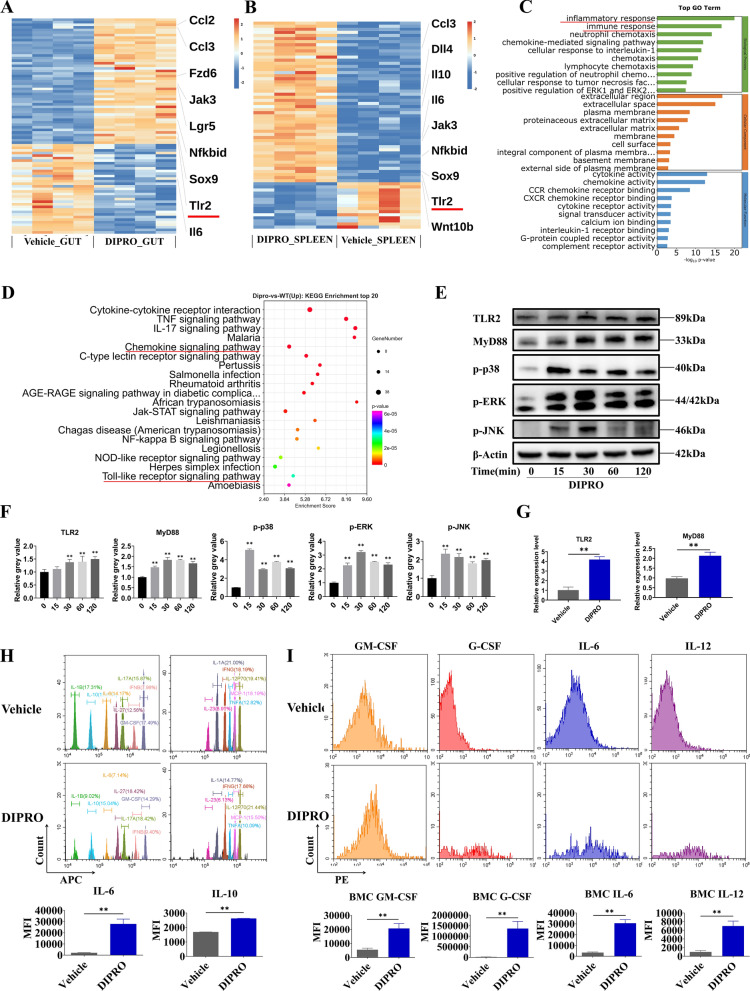
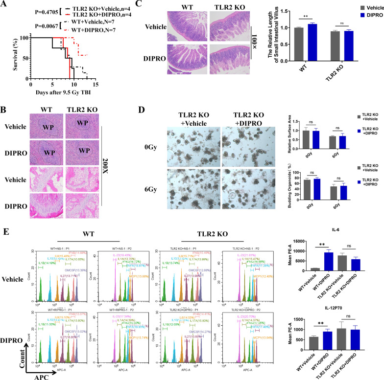
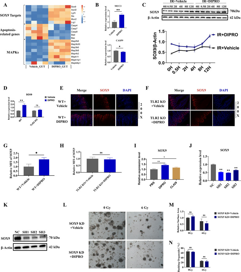
Severe ionizing radiation (IR) causes the acute lethal damage of hematopoietic system and gastrointestinal tract. By establishing a radiation injury model, we found that Diprovocim, a TLR2 agonist, protected mice against the lethal damage of hematopoietic system and gastrointestinal tract. Diprovocim inhibited the IR-induced damage, promoted erythrocyte differentiation and elevated the proportion of hematopoietic stem cells (HSCs) in irradiated mice, and promoted the proliferation and differentiation of intestinal stem cells (ISCs). In addition, the RNA seq results suggested that Diprovocim significantly upregulated the TLR2 signaling pathway, and Diprovocim had no radioprotective effect on TLR2 KO mice, suggesting that Diprovocim activated TLR2 signaling pathway to exert its radioprotective function. The RNA sequencing results also suggested that Diprovocim significantly up-regulated the expression of SOX9. Diprovocim had no radioprotective effect after SOX9 knockdown. In conclusion, we demonstrated that Diprovocim protected the radiation-induced damage and upregulated targeting TLR2-SOX9 axis and that Diprovocim might be a potential high-efficiency selective agent.

Keywords: Diprovocim; Intestinal stem cells; Ionizing radiation-induced injury; SOX9; TLR2.


Conflict of interest statement

Declarations. Ethics approval and consent to participate: All animal experiments conformed to the National Institute of Health Guide for the Care and Use of Laboratory Animals'(NIH Publication No. 85–23, National Academy Press, Washington, DC, revised 1996), with the approval of the Laboratory Animal Center of the Naval Medical University, Shanghai. Consent for publication: Written informed consent for publication was obtained from all participants. Competing interests: The authors declare no competing interests.

Figures

Fig. 1
Fig. 1
Diprovocim significantly ameliorated IR-induced damage. A, B Male mice injected intraperitoneally with Diprovocim at 18 h and 2 h before IR, and then exposed to 7.5 Gy, 12.0 Gy TBI. Control mice were treated with Normal Saline. Survival was recorded. C The change of body weight from Day 0 to Day 5 in each group of mice after 12 Gy TBI (n = 3). D Mice were treated with vehicle or Diprovocim, irradiated at 12 Gy for 1 day, and the count of WBC was determined by using blood cell analyzer (n = 3). E Male mice were injected intraperitoneally with Diprovocim at 18 h and 2 h after 9.5 Gy TBI. Control mice were treated with Normal Saline. Survival was recorded. F MODE-K cells were stimulated with Diprovocim (0.5 μM) 2 h after 8 Gy IR, and cell viability was determined by CCK-8 (n = 6). The data were presented as mean ± SD. *p < 0.05, **p < 0.01, ns indicates no statistical significance
Fig. 2
Fig. 2
Diprovocim protected the intestinal tissue against radiation-induced injury. A Representative images of HE-stained intestinal sections at 3.5 day after 9.5 Gy TBI (n = 3). B Villus length (n = 3). C The representative images of OLFM4 immunofluorescences intestinal sections at 3.5 days after 9.5 Gy TBI (n = 3). D The relative MFI of OLFM4. E qRT-PCR results of Lgr5, OLFM4 and ASCL2 (n = 3). F The radioprotection of Diprovocim on MODE-K cells viability as determined by CCK-8 assay (n = 6). G Intestinal crypts of C57BL/6 mice were extracted for organoid culture, and then it was stimulated with Diprovocim or vehicle. H The relative area of intestinal organoids and the percent of budding intestinal organoids. The data were presented as mean ± SD. *p < 0.05, **p < 0.01, ns indicates no statistical significance
Fig. 3
Fig. 3
Diprovocim protected the hematopoietic system against radiation-induced injury. A Representative images of HE-stained spleen and bone marrow sections after 1 d,14 d, and 28 d of 8.0 Gy IR (n = 3). BG Bone marrow cells (BMCs) in mice were isolated at 24 h after 8.0 Gy TBI for flow cytometry analysis (n = 3). B The proportions of LK, LSK ratio was detected by using flow cytometry. C The proportions of pre-mature B cells, immature B cells and mature B cells was detected by using flow cytometry. D The proportion of Pro.E, Ery.A, Ery.B, Ery.C was detected by using flow cytometry. E The proportion of MDSC was detected by using flow cytometry. F The proportion of macrophage was detected by using flow cytometry. G BMCs apoptosis was detected by using flow cytometry (n = 3). The data were presented as mean ± SD. *p < 0.05, **p < 0.01, ns indicates no statistical significance
Fig. 4
Fig. 4
Diprovocim exerted radioprotection through the TLR2 signaling pathway. A, B Heatmap of DEGs between IR + vehicle mice and IR + Diprovocim mice (n = 4). C GO term analysis was performed on DEGs. D Pathway enrichment analysis of KEGG. E Immunoblot analysis of lysates of MODE-K with 0.5 μM Diprovocim at the indicated times. F The quantitative analysis of protein. G The expression of TLR2 and MyD88 in intestinal tissue with vehicle or Diprovocim treatment (n = 3). H The levels of IL-6 and IL-10 in mouse serum (n = 3). I Mean fluorescence intensity of GM-CSF, G-CSF, IL-6 and IL-12 was measured by flow cytometry (n = 3). The data were presented as mean ± SD. *p < 0.05, **p < 0.01
Fig. 5
Fig. 5
The radioprotection of Diprovocim was TLR2 dependent. A C57BL/6 mice and TLR2 KO mice were intraperitoneally injected with vehicle or Diprovocim before 9.5 Gy TBI. Survival was recorded. B Representative HE staining images of spleen and bone marrow sections at 3.5 days after 9.5 Gy TBI (n = 3). C Representative images of HE-stained intestinal sections after 3.5 days of 9.5 Gy TBI and villus length (n = 3). D Intestinal crypts of TLR2 KO mice were extracted for organoid culture, and then it was stimulated with Diprovocim or vehicle. E Cytokine changes in serum at day 1 after 9.5 Gy TBI (n = 3). The data were presented as mean ± SD. *p < 0.05, **p < 0.01, ns indicates no statistical significance
Fig. 6
Fig. 6
The intestinal radioprotection of Diprovocim was dependent on SOX9. A The DEGs of SOX9 downstream target genes, apoptosis-related genes, and MAPK signaling bteween IR + vehicle mice and IR + Diprovocim mice (n = 4). B qRT-PCR results of MUC2 and CASP9 in the intestine at 3.5 days after 9.5 Gy TBI (n = 3). C WB analysis of MODE-K cells proteins. D qRT-PCR results of SOX9 in intestinal tissue of C57BL/6 mice and TLR2 KO mice with vehicle or Diprovocim treatment at 3.5 days after 9.5 Gy TBI (n = 3). E The representative images of SOX9 immunofluorescences in WT mice at 3.5 days after 9.5 Gy TBI (n = 3). F The representative images of SOX9 immunofluorescences in TLR2 KO mice at 3.5 days after 9.5 Gy TBI (n = 3). G The relative MFI of SOX9 in WT mice. H The relative MFI of SOX9 in TLR2 KO mice. I The expression of SOX9 in MODE-K cells after treatment with the Diprovocim (0.5 μM) or CL429 (2 μg/mL). J The relative expression of SOX9 after shRNA knockdown. K The validation of SOX9 knockdown at the protein level. L Intestinal crypts of C57BL/6 mice were extracted and used to establish SOX9 KD intestinal organoids, and then it was stimulated with Diprovocim or vehicle. M Relative surface of intestinal organoids. N Budding rate of intestinal organoids. The data were presented as mean ± SD. *p < 0.05, **p < 0.01, ns indicates no statistical significance

Similar articles

References

    1. Akira S, Uematsu S, Takeuchi O. Pathogen recognition and innate immunity. Cell. 2006;124:783–801. 10.1016/j.cell.2006.02.015. - PubMed
    1. Akiyama H, Kim JE, Nakashima K, Balmes G, Iwai N, Deng JM, Zhang Z, Martin JF, Behringer RR, Nakamura T, de Crombrugghe B. Osteo-chondroprogenitor cells are derived from Sox9 expressing precursors. Proc Natl Acad Sci U S A. 2005;102:14665–70. 10.1073/pnas.0504750102. - PMC - PubMed
    1. Bell BI, Koduri S, Salas Salinas C, Monslow J, Puré E, Ben-Josef E, Koumenis C, Verginadis II. Interleukin 6 signaling blockade exacerbates acute and late injury from focal intestinal irradiation. Int J Radiat Oncol Biol Phys. 2019;103:719–27. 10.1016/j.ijrobp.2018.10.007. - PMC - PubMed
    1. Bergstrom KS, Kissoon-Singh V, Gibson DL, Ma C, Montero M, Sham HP, Ryz N, Huang T, Velcich A, Finlay BB, et al. Muc2 protects against lethal infectious colitis by disassociating pathogenic and commensal bacteria from the colonic mucosa. PLoS Pathog. 2010;6:e1000902. 10.1371/journal.ppat.1000902. - PMC - PubMed
    1. Brickey WJ, Caudell DL, Macintyre AN, Olson JD, Dai Y, Li S, Dugan GO, Bourland JD, O’Donnell LM, Tooze JA, et al. The TLR2/TLR6 ligand FSL-1 mitigates radiation-induced hematopoietic injury in mice and nonhuman primates. Proc Natl Acad Sci U S A. 2023;120:e2122178120. 10.1073/pnas.2122178120. - PMC - PubMed

MeSH terms

LinkOut - more resources