

Dynamic evolution and antitumor mechanisms of CXCR6+CD8+ T cells in small cell lung cancer treated with low-dose radiotherapy and immunotherapy
- PMID: 40247265
- PMCID: PMC12007177
- DOI: 10.1186/s12967-025-06450-1
Dynamic evolution and antitumor mechanisms of CXCR6+CD8+ T cells in small cell lung cancer treated with low-dose radiotherapy and immunotherapy
Abstract
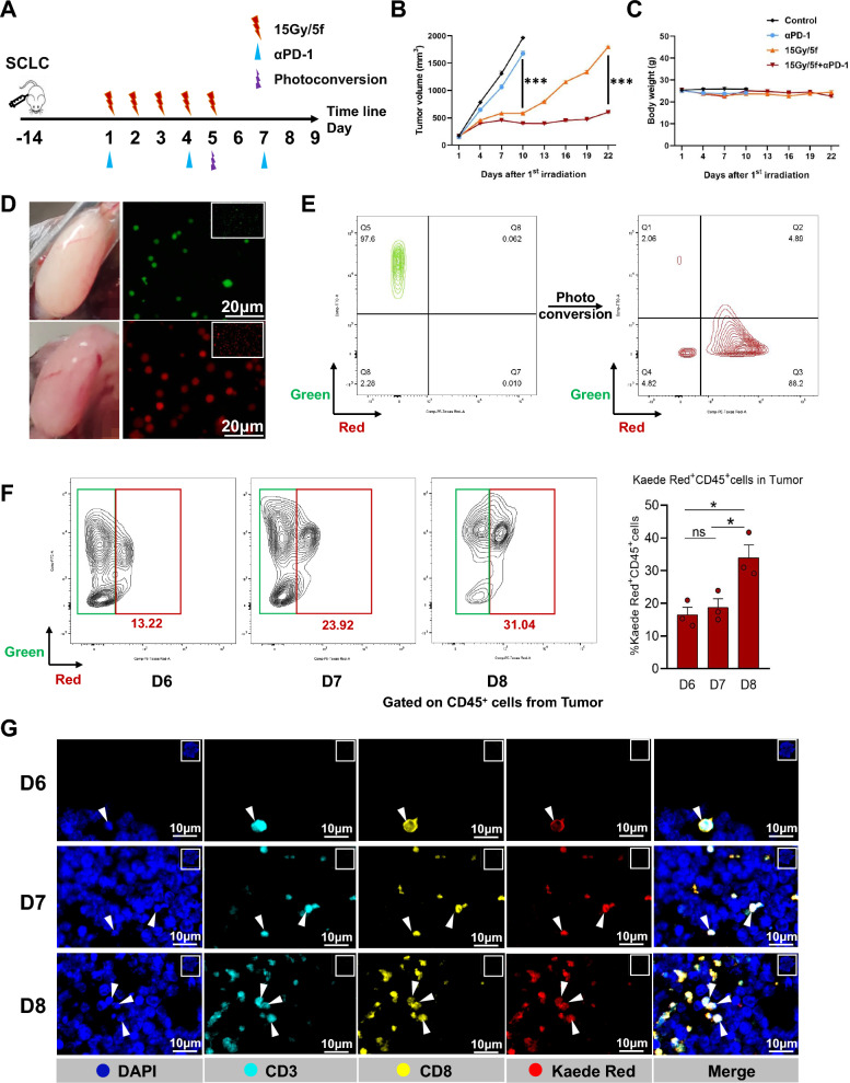
Background: Patients with small-cell lung cancer (SCLC) have the poor prognosis. Current research suggested that low-dose radiotherapy (LDRT) combined with immunotherapy can enhance the immunogenicity of tumor cells, thereby improving antigen presentation and promoting the intratumoral infiltration of CD8+ T cells, which significantly extends the survival of patients. However, the change trajectory of T cells, and the mechanisms underlying the promotion of intratumoral infiltration of CD8+ T cells, and the enhancement of their cytotoxic functions remain to be elucidated.
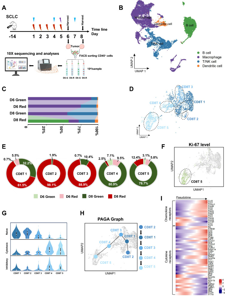
Methods: To delineate the dynamic changes of T cells, we collected tumors from Kaede tumor-bearing mice that had undergone radioimmunotherapy. Using flow cytometry, we sorted intratumoral-infiltrating immune cells, which were required for single-cell RNA sequencing, at various time points (Kaede Red: derived from tumor-draining lymph node [TDLN]). The results obtained from the sequencing analysis were further validated through experiments, such as flow cytometry, immunofluorescence, and analysis of clinical cohort data.
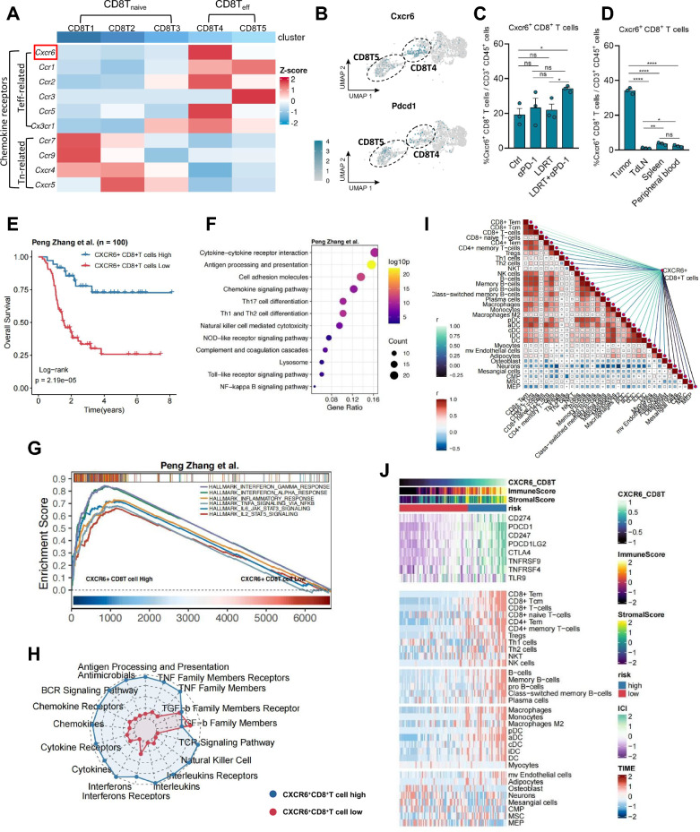
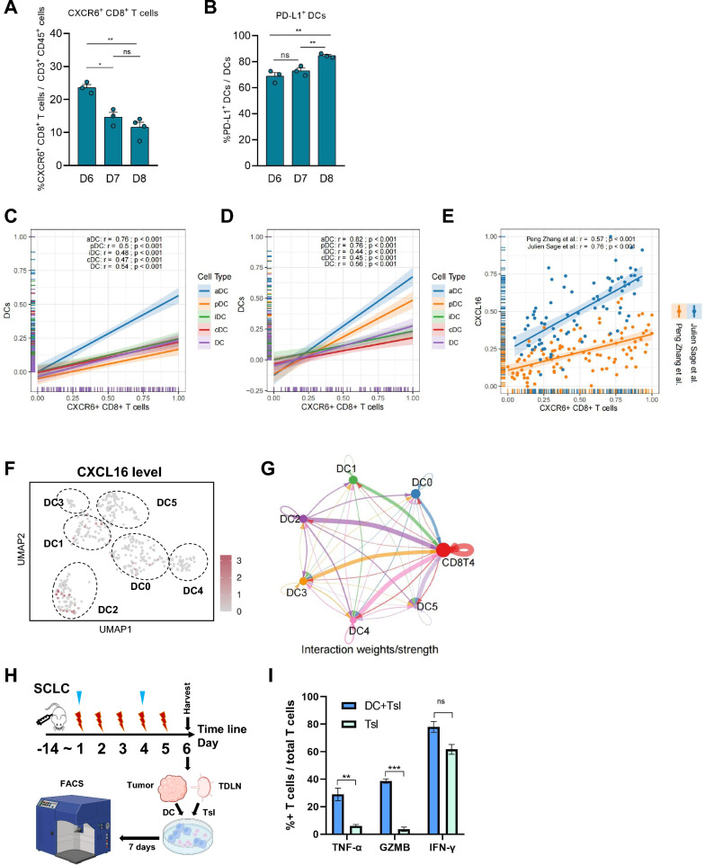
Results: Here, we observed stem-like T cells migrating from the TDLN to the tumor site and differentiating into effector phenotypes within the tumor. Dendritic cells (DCs) are the key cluster that induces the differentiation of stem-like T cell into effector phenotypes. Moreover, SCLC patients with a high infiltration of tumor-specific CXCR6+CD8+ T cells exhibited a supportive TME and longer survival time (P < 0.001).
Conclusions: This study delineates the change trajectory of CD8+ T cells, identifies the crucial role of DCs in T cell differentiation, and highlights the significance of tumor-specific CXCR6+CD8+ T cells in anti-tumor immunity. Future therapeutic strategies for SCLC could focus on enhancing the infiltration of activated DCs and CXCR6+CD8+ T cells within the tumor microenvironment to improve treatment efficacy.
Keywords: CD8-positive T-lymphocytes; Immunotherapy; Radiotherapy; Small-cell lung cancer; Tumor-infiltrating.
© 2025. The Author(s).
Conflict of interest statement
Declarations. Ethics approval and consent to participate: Our study was approved by Animal Ethics Committee of West China Hospital, Sichuan University (Ethics Approval Number: 20230306082), the animal experiments involved in this study have adhered to ARRIVE guidelines. Consent for publication: All authors have read and agreed to the published version of the manuscript. Competing interests: The authors have no conflicts of interest to declare.
Figures
Similar articles
-
In situ delivery of iPSC-derived dendritic cells with local radiotherapy generates systemic antitumor immunity and potentiates PD-L1 blockade in preclinical poorly immunogenic tumor models.J Immunother Cancer. 2021 May;9(5):e002432. doi: 10.1136/jitc-2021-002432. J Immunother Cancer. 2021. PMID: 34049930 Free PMC article.
-
CXCR6 deficiency impairs cancer vaccine efficacy and CD8+ resident memory T-cell recruitment in head and neck and lung tumors.J Immunother Cancer. 2021 Mar;9(3):e001948. doi: 10.1136/jitc-2020-001948. J Immunother Cancer. 2021. PMID: 33692218 Free PMC article.
-
CXCR6 is required for antitumor efficacy of intratumoral CD8+ T cell.J Immunother Cancer. 2021 Aug;9(8):e003100. doi: 10.1136/jitc-2021-003100. J Immunother Cancer. 2021. PMID: 34462326 Free PMC article.
-
Combined treatment of small cell lung cancer using radiotherapy and immunotherapy: Challenges and updates.Biomed Pharmacother. 2025 Jan;182:117727. doi: 10.1016/j.biopha.2024.117727. Epub 2024 Dec 14. Biomed Pharmacother. 2025. PMID: 39675137 Review.
-
Tumor-induced CD8+ T-cell dysfunction in lung cancer patients.Clin Dev Immunol. 2012;2012:741741. doi: 10.1155/2012/741741. Epub 2012 Oct 17. Clin Dev Immunol. 2012. PMID: 23118782 Free PMC article. Review.
References
-
- Abu Rous F, Singhi EK, Sridhar A, Faisal MS, Desai A. Lung cancer treatment advances in 2022. Cancer Invest. 2023;41(1):12–24. - PubMed
-
- Tran KB, Lang JJ, Compton K, Xu R, Acheson AR, Henrikson HJ, Kocarnik JM, Penberthy L, Aali A, Abbas Q, Abbasi B. The global burden of cancer attributable to risk factors, 2010–19: a systematic analysis for the Global Burden of Disease Study 2019. Lancet (London, England). 2022;400(10352):563–91. - PMC - PubMed
-
- Lee JH, Saxena A, Giaccone G. Advancements in small cell lung cancer. Semin Cancer Biol. 2023;93:123–8. - PubMed
-
- Caliman E, Fancelli S, Petroni G, Gatta Michelet MR, Cosso F, Ottanelli C, Mazzoni F, Voltolini L, Pillozzi S, Antonuzzo L. Challenges in the treatment of small cell lung cancer in the era of immunotherapy and molecular classification. Lung cancer (Amsterdam Netherlands). 2023;175:88–100. - PubMed
-
- Yu WD, Sun G, Li J, Xu J, Wang X. Mechanisms and therapeutic potentials of cancer immunotherapy in combination with radiotherapy and/or chemotherapy. Cancer Lett. 2019;452:66–70. - PubMed
MeSH terms
Substances
Grants and funding
LinkOut - more resources
Full Text Sources
Medical
Research Materials
