

Visual and anatomical outcomes of primary retinectomy for diabetic tractional retinal detachment
- PMID: 40247269
- PMCID: PMC12004879
- DOI: 10.1186/s12886-025-04065-0
Visual and anatomical outcomes of primary retinectomy for diabetic tractional retinal detachment
Abstract
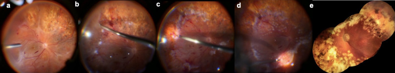
Purpose: Uncontrolled proliferative diabetic retinopathy (PDR) can cause fibrovascular growth and retinal traction, leading to tractional retinal detachment (TRD). The role of primary retinectomy in diabetic TRD remains unclear, as most studies focus on rhegmatogenous retinal detachment (RRD) with PVR. This study aims to investigate the impact of retinectomy on anatomical and visual outcomes in patients undergoing pars plana vitrectomy (PPV) for diabetic TRD.
Method: Patients who underwent primary retinectomy during PPV for diabetic TRD were retrospectively evaluated. Best corrected visual acuity (BCVA) before surgery and at the final follow-up, retinectomy characteristics, and final retinal attachment status were documented. TRD score, the quadrant and extent of the retinectomy, presence of macular displacement at final follow-up, and postoperative complications were evaluated. The relationship between the quadrants and extent of the retinectomy and visual acuity was also assessed.
Result: Thirty-eight eyes of 38 patients with mean age 60.55 ± 10.00 years were included. Mean follow-up was 23.53 ± 27.40 months. The most common locations of the retinectomy sites were extended posterior to the equator (39.5%), around the equatorial zone (34.2%), and peripheral retina (26.3%). The mean BCVA improved from 1.71 ± 0.53 logMAR to 1.48 ± 0.74 logMAR at the final follow-up. At the final visit 65.8% of patients experienced improved or maintained BCVA. Temporal retinectomy showed worse visual outcomes in the Chi-square test but not in binary logistic regression analysis. Furthermore, 26 (68.4%) eyes were attached without tamponade, 10 (26.3%) were attached under silicone oil and 2 (5.6%) remained detached under silicone oil.
Conclusion: These findings suggest that retinectomy, when deemed necessary in eyes with diabetic TRD, may not lead to poor functional and anatomical outcomes, contrary to some previous assumptions.
Keywords: Proliferative diabetic retinopathy; Retinectomy; Tractional retinal detachment.
© 2025. The Author(s).
Conflict of interest statement
Declarations. Ethical approval: This investigation complied with the principles outlined in the Declaration of Helsinki. The local ethics committee approved the study (Number: GOKAEK-2024/07.06). Written informed consent was obtained from all participants before participating. Competing interests: The authors declare no competing interests.
Figures

Similar articles
-
Macular changes after primary retinectomy for retinal detachment complicated by proliferative vitreoretinopathy.Clin Exp Optom. 2024 May;107(4):434-441. doi: 10.1080/08164622.2023.2236098. Epub 2023 Sep 6. Clin Exp Optom. 2024. PMID: 37674262
-
Outcomes, efficacy and risk factors of 27-Gauge vitrectomy for diabetic tractional retinal detachment in Japanese patients.Jpn J Ophthalmol. 2025 Jan;69(1):59-65. doi: 10.1007/s10384-024-01135-6. Epub 2024 Nov 6. Jpn J Ophthalmol. 2025. PMID: 39503819 Free PMC article.
-
Evaluation of Intravitreal Ranibizumab on the Surgical Outcome for Diabetic Retinopathy With Tractional Retinal Detachment.Medicine (Baltimore). 2016 Feb;95(8):e2731. doi: 10.1097/MD.0000000000002731. Medicine (Baltimore). 2016. PMID: 26937902 Free PMC article.
-
Surgical management of retinal diseases: proliferative diabetic retinopathy and traction retinal detachment.Dev Ophthalmol. 2014;54:196-203. doi: 10.1159/000360467. Epub 2014 Aug 26. Dev Ophthalmol. 2014. PMID: 25196770 Review.
-
Predicting visual outcome following retinectomy for retinal detachment.Br J Ophthalmol. 2008 Jul;92(7):954-8. doi: 10.1136/bjo.2007.131540. Epub 2008 Jun 12. Br J Ophthalmol. 2008. PMID: 18556423 Review.
References
-
- Congdon NG, Friedman DS, Lietman T. Important causes of visual impairment in the world today. JAMA. 2003;290(15):2057–60. - PubMed
-
- Diabetic Retinopathy Study Research Group. Four risk factors for severe visual loss in diabetic retinopathy: the third report from the diabetic retinopathy study. Arch Ophthalmol. 1979;97(4):654–5. - PubMed
-
- Kroll P, Rodrigues EB, Hoerle S. Pathogenesis and classification of proliferative diabetic vitreoretinopathy. Ophthalmologica. 2007;221(2):78–94. - PubMed
-
- Simó R, Carrasco E, García-Ramírez M, Hernández C. Angiogenic and antiangiogenic factors in proliferative diabetic retinopathy. Curr Diabetes Rev. 2006;2(1):71–98. - PubMed
-
- Eliott D, Hemeida T. Diabetic traction retinal detachment. Int Ophthalmol Clin. 2009;49(2):153–65. - PubMed
MeSH terms
LinkOut - more resources
Full Text Sources
Medical
Research Materials
Miscellaneous
