

The rolling stone: migration of an intrauterine device leading to bladder stone formation nine years after insertion: a case report
- PMID: 40247326
- PMCID: PMC12004547
- DOI: 10.1186/s12894-025-01780-0
The rolling stone: migration of an intrauterine device leading to bladder stone formation nine years after insertion: a case report
Abstract
Background: Intrauterine devices are safe, affordable, convenient, and the most common form of contraception used by females of childbearing age in Palestine. A rare complication of intrauterine devices is migration to nearby structures, rarely the urinary bladder, leading to bladder stone formation.
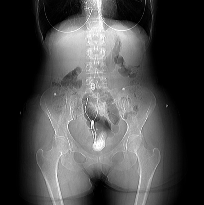
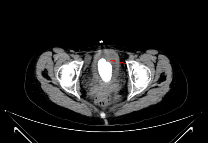
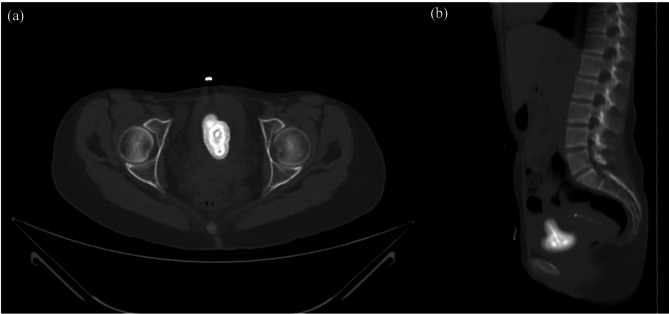
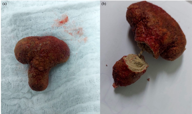
Case presentation: A 34-year-old female patient presented due to repeated urinary tract infections and flank pain associated with lower urinary tract symptoms, including dysuria, frequency, and gross hematuria. Subsequent laboratory tests revealed a past medical history of iron-deficiency anemia. Urinalysis revealed hematuria and pyuria, and the urine culture confirmed colonization of Escherichia coli. Computed tomography revealed an irregularly shaped 5.5 cm hyperdense calculus in the urinary bladder. Open cystolithotomy was done to extract the calculus, which was later incidentally revealed to be encrusting a migrated intrauterine device.
Conclusions: This case highlights the rare potential for intrauterine devices to migrate to the urinary bladder, leading to calculus formation, which, in this case, was discovered in this patient nine years post-insertion. The intrauterine device perforation into the urinary bladder was due to delayed inflammatory migration. This case underscores the critical need for both patient and physician education in low-resource settings on the warning signs of intrauterine device migration, including new-onset irritative lower urinary tract symptoms, hematuria, and missing intrauterine device threads, ensuring routine scheduled follow-ups, patient self-checks, and timely imaging can aid in early detection and prevent complications associated with intrauterine device migration.
Keywords: Copper IUD; IUD migration; Lower urinary tract symptoms; Open cystolithotomy; Recurrent urinary tract infections; Urinary bladder stone.
© 2025. The Author(s).
Conflict of interest statement
Declarations. Ethical approval and consent: This case report was approved by the Al-Quds University ethical committee and informed written consent was obtained from the patient herself. All methods utilized in this case were conducted following the relevant regulations and guidelines in accordance to the principles of the World Medical Association (WMA) Declaration of Helsinki. Consent to publish: The authors declare that written informed consent was obtained from the patient for the publication of this manuscript and any accompanying figures. Competing interests: The authors declare no competing interests.
Figures
References
-
- United Nations Department of Economic and Social Affairs, Population Division. World Family Planning 2022: Meeting the changing needs for family planning: Contraceptive use by age and method. 2022. Available from: UN DESA/POP/2022/TR/NO. 4.
Publication types
MeSH terms
LinkOut - more resources
Full Text Sources
Miscellaneous
