. 2025 Apr 17;30(1):296.
doi: 10.1186/s40001-025-02557-9.

Mammalian Ste20-like kinase 1 regulates AMPK to mitigate the progression of non-alcoholic fatty liver disease

Affiliations

Mammalian Ste20-like kinase 1 regulates AMPK to mitigate the progression of non-alcoholic fatty liver disease

Lijuan Wang et al. Eur J Med Res. .

Abstract

Background: Non-alcoholic steatohepatitis (NASH) progression is strongly associated with deteriorating hepatic function, primarily driven by free cholesterol (FC) accumulation-induced lipotoxicity. Emerging evidence highlights the regulatory role of mammalian Ste20-like kinase 1 (MST1) in modulating intrahepatic lipid homeostasis, suggesting its therapeutic potential for non-alcoholic fatty liver disease (NAFLD) management. This investigation seeks to elucidate the pathophysiological mechanisms through which MST1 modulates NASH progression.

Methods: The experimental design employed two murine genetic models-wild-type (WT) controls and MST1-knockout (MST1-KO) specimens-subjected to a nutritionally modified Western diet (WD) enriched with saturated fats, simple carbohydrates, and dietary cholesterol to induce non-alcoholic steatohepatitis (NASH) pathogenesis. Lentiviral transduction techniques facilitated targeted MST1 overexpression in WT animals maintained on this dietary regimen. Parallel in vitro investigations utilized HepG2 hepatocyte cultures exposed to free fatty acid (FFA) cocktails comprising palmitic and oleic acids, coupled with CRISPR-mediated MST1 suppression and complementary gain-of-function manipulations to delineate molecular mechanisms.

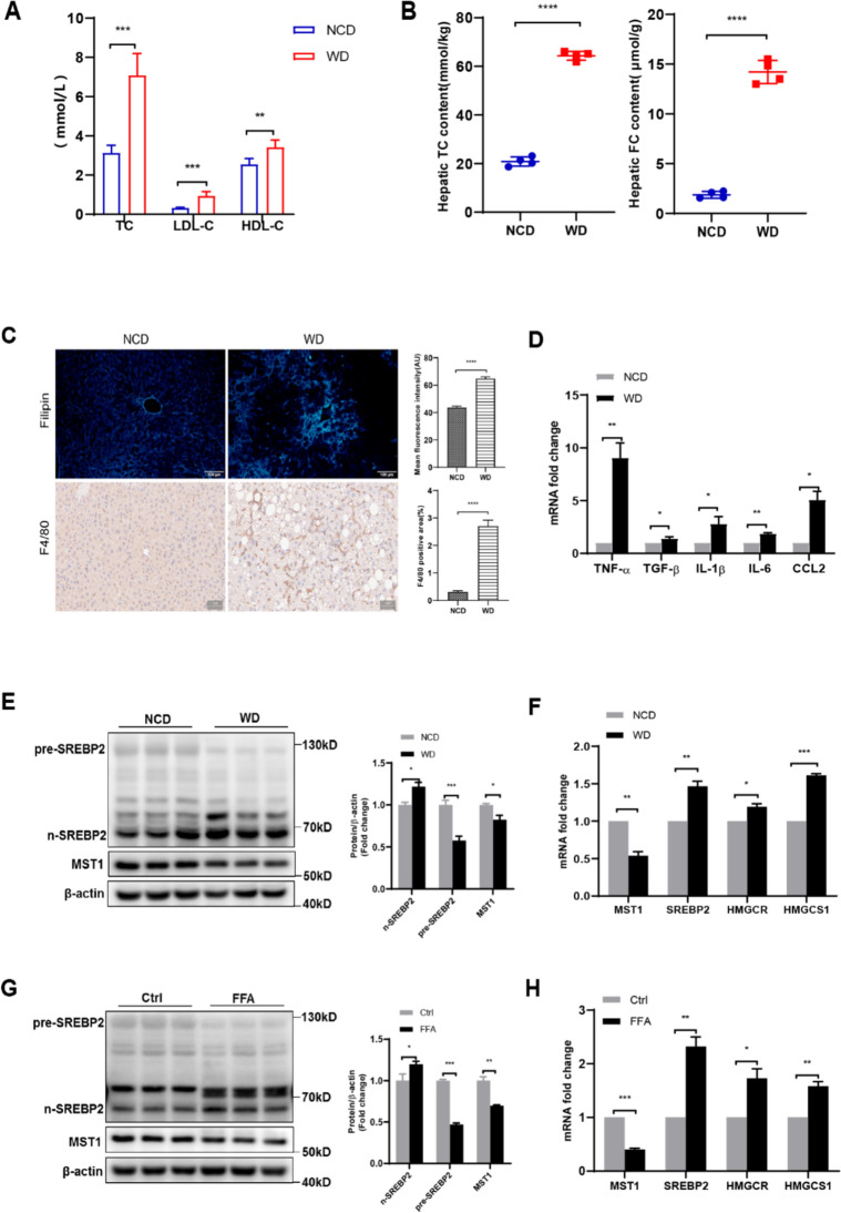
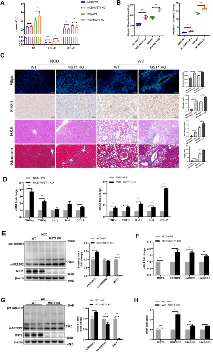
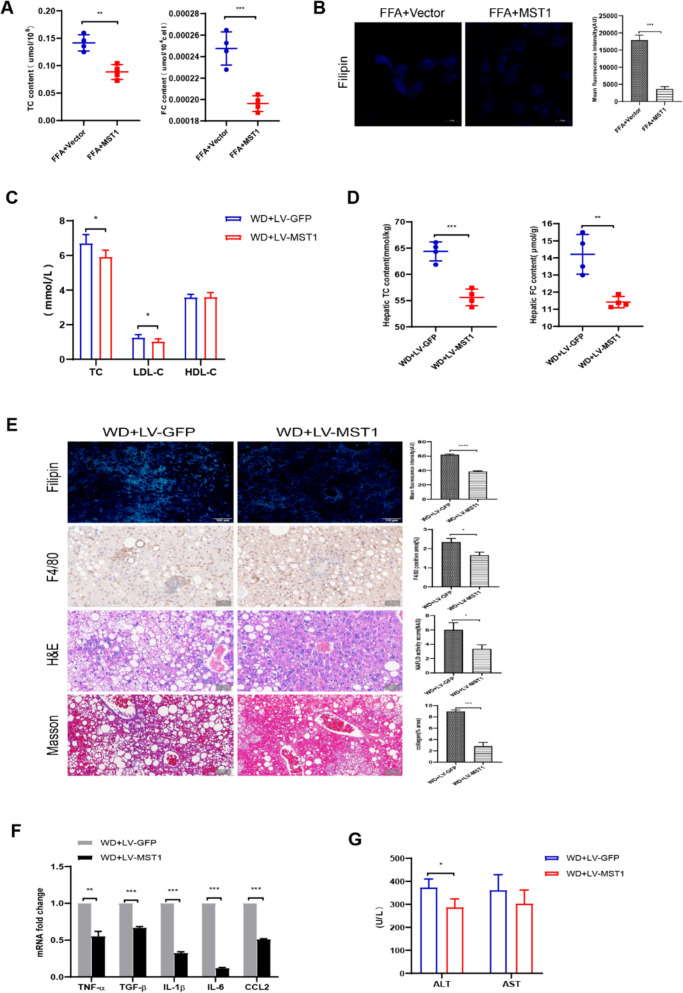
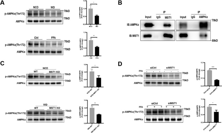
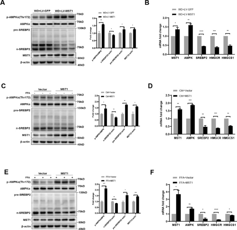
Results: NASH triggers hepatic sterol biosynthesis activation, resulting in pathological FC overload concurrent with MST1 transcriptional suppression. Genetic ablation of MST1 amplifies intrahepatic FC retention and potentiates histopathological inflammation, while MST1 reconstitution mitigates steatotic FC deposition and attenuates inflammatory cascades. Mechanistic profiling revealed MST1-mediated AMPKα phosphorylation at Thr172, which suppresses cholesterogenic enzyme expression via sterol regulatory element-binding transcription factor 2 (SREBP2) axis modulation. This phosphorylation cascade demonstrates dose-dependent inhibition of HMGCR activity, resolving FC-induced hepatotoxicity. Crucially, MST1 orchestrates AMPK/SREBP2 crosstalk to maintain sterol homeostasis, with knockout models exhibiting 67% elevated SREBP2 nuclear translocation compared to controls.

Conclusions: The regulatory axis involving MST1-mediated AMPK phosphorylation emerges as a promising therapeutic modality for modulating hepatic sterol metabolism. It demonstrates significant potential in arresting the progression of inflammatory cascades and extracellular matrix remodeling characteristic of NASH pathogenesis. Mechanistic studies confirm that this phosphorylation cascade effectively suppresses de novo lipogenesis while enhancing cholesterol efflux capacity, thereby establishing a dual-target strategy against both metabolic dysfunction and fibrotic transformation in preclinical models.

Keywords: AMP-activated protein kinase; Cholesterol synthesis; Hepatic free cholesterol; Mammalian sterile 20-like kinase 1; Non-alcoholic steatohepatitis.


Conflict of interest statement

Declarations. Ethics approval and consent to participate: This study strictly followed the Guidelines for the Care and Use of Experimental Animals published by the National Institutes of Health, and all experimental protocols were approved by the Medical Ethics Review Committee of Ningxia Medical University (IACUC- 2023 - 001). Competing interests: The authors declare no competing interests.

Figures

Fig. 1
Fig. 1
NASH-associated hepatic cholesterol dysregulation correlates with activated cholesterol biosynthetic signaling and attenuated MST1 transcriptional activity. C57BL6/J mice received NCD/WD dietary regimens for 18 weeks with subsequent analyses: a circulating lipid profile quantification including TC, LDL-cholesterol, and HDL-C concentrations. b Hepatic compartmentalization of esterified versus FC pools. c Histomorphometric characterization through Filipin-stained cholesterol microdomains (100 ×) and F4/80 + macrophage infiltration mapping (200 ×), with quantitative fluorescence intensity and cellular infiltration area measurements. d Transcriptional activation patterns of inflammatory mediators and extracellular matrix remodeling factors in hepatic tissue. e Immunoblot analysis with densitometric quantification of SREBP2 proteolytic processing and MST1 expression profiles in murine liver specimens. f Hepatic transcriptional landscape of cholesterol regulatory network components. g FFA-challenged HepG2 cellular models demonstrating SREBP2 maturation dynamics and MST1 expression through immunoblot quantification. h Lipid-induced transcriptional reprogramming of cholesterol biosynthesis machinery in hepatocyte models. Data expressed as mean ± SEM (n = 6/group) from triplicate experimental replicates* versus control cohorts, with statistical significance thresholds defined as *P < 0.05, **P < 0.01, ***P < 0.001, ****P < 0.0001
Fig. 2
Fig. 2
MST1 deficiency disrupts hepatic cholesterol homeostasis, potentiates lipotoxic injury and inflammatory cascades, while activating cholesterol biosynthetic pathways. MST1-KO murine models subjected to 18-week NCD/WD dietary interventions exhibited: a serum lipid profile alterations including TC, LDL-cholesterol, and HDL-C concentrations. b Quantitative assessment of hepatic total and FC accumulation. c Histomorphometric characterization through Filipin-stained cholesterol visualization (100x), F4/80 + macrophage infiltration mapping, H&E-stained parenchymal architecture, and collagen deposition analysis via Masson’s trichrome (200x), with quantitative metrics for fluorescent intensity, inflammatory cell distribution, fibrotic content, and NAFLD pathological scoring. d Transcriptional activation patterns of inflammatory mediators and fibrogenesis markers in hepatic tissue. e Immunoblot analysis with densitometric quantification of SREBP2 proteolytic processing and MST1 expression in NCD-fed liver specimens. f mRNA profiling of cholesterol regulatory network components in NCD-treated hepatic samples. g Western blot densitometry illustrating SREBP2 maturation dynamics and MST1 levels in WD-exposed liver tissues. h WD-induced transcriptional reprogramming of cholesterol biosynthetic machinery components. Data expressed as mean ± SEM (n = 6/group) from triplicate experimental replicates* versus respective controls, with statistical significance denoted as *P < 0.05, **P < 0.01, ***P < 0.001, ****P < 0.0001
Fig. 3
Fig. 3
Impact of MST1 overexpression on Cholesterol Metabolism, inflammatory response, and hepatic injury. a Effect of MST1 overexpression on total cholesterol (TC) and free cholesterol (FC) levels in FFA-stimulated HepG2 cells. b Filipin staining showing membrane FC distribution patterns in FFA-stimulated HepG2 cells. Bar graphs represent quantitative analysis of mean fluorescence intensity. c Influence of MST1 overexpression on serum TC, LDL-C (low-density lipoprotein cholesterol), and HDL-C (high-density lipoprotein cholesterol) concentrations in Western diet (WD)-fed mice. d Effect of MST1 overexpression on hepatic and plasma FC levels in WD-fed mice. e Liver tissue staining in WD-fed mice, including Filipin (FC distribution), F4/80 (macrophage marker), H&E (histomorphology), Masson (fibrosis), and corresponding quantitative analyses. f qPCR analysis of relative expression changes in TNF-α, TGF-β, IL-1β, IL-6, and CCL2. g Measurement of alanine aminotransferase (ALT) and aspartate aminotransferase (AST) levels following MST1 overexpression. Data expressed as mean ± SEM from triplicate independent experiments. Statistical significance denoted as *p < 0.05, **p < 0.01, ***p < 0.001, ****p < 0.0001 versus corresponding control groups
Fig. 4
Fig. 4
MST1 mediates Thr172 phosphorylation of AMPKα. a Western blot analysis with densitometric quantification of p-AMPKα and AMPKα expression in hepatic tissues from in vivo and in vitro NASH models. b Co-immunoprecipitation assays performed using MST1-specific and AMPK-specific antibodies to demonstrate protein–protein interactions. c Immunoblot quantification of AMPK phosphorylation status in liver samples from MST1-deficient mice under NCD and WD dietary regimens. d Phosphorylation profile analysis through immunoblotting and densitometry in FFA-stimulated versus basal HepG2 cells following MST1 silencing. Data expressed as mean ± SEM from triplicate independent experiments. Statistical significance denoted as *P < 0.05, **P < 0.01, ***P < 0.001, ****P < 0.0001 versus corresponding control groups
Fig. 5
Fig. 5
AMPK activation counteracts hepatic cholesterol deposition and MST1 deficiency-induced hepatotoxicity. MST1-deficient murine models receiving NCD/WD regimens for 16 weeks underwent subsequent 14-day AICAR administration via intraperitoneal route. a Serum lipid profiles including TC, LDL-C, and HDL-C concentrations. b Hepatic quantification of TC and FC levels. c Histopathological characterization through Filipin-stained fluorescence microscopy (100x), F4/80 + macrophage infiltration, hematoxylin–eosin stained parenchyma, and collagen deposition visualized by Masson’s trichrome (200x), with quantitative morphometric analysis of fluorescence intensity, macrophage distribution area, fibrotic content, and NAFLD pathological scoring. d Transcriptional activation profiles of inflammatory mediators and fibrogenesis markers in hepatic tissue. e Immunoblot analysis with densitometric quantification of AMPK phosphorylation status, SREBP2 proteolytic processing, and MST1 expression in NCD-fed murine livers. f mRNA profiling of AMPK signaling components and cholesterol biosynthesis enzymes in NCD-treated hepatic samples. g Immunoblot densitometry illustrating AMPK pathway activation dynamics and SREBP2 maturation in WD-exposed liver specimens. h WD-challenged hepatic transcriptome alterations in cholesterol regulatory network components. Data expressed as mean ± SEM from sextuplicate biological replicates across three experimental iterations* versus respective controls. *P < 0.05, **P < 0.01, ***P < 0.001, ****P < 0.0001
Fig. 6
Fig. 6
MST1 regulates cholesterol biosynthesis via modulation of the AMPK/SREBP2 axis. a Western blot analysis with densitometric quantification demonstrated hepatic expression profiles of p-AMPKα, AMPKα, nuclear SREBP2, precursor SREBP2, and MST1 in WD-fed C57BL6/J mice, including successful MST1 overexpression mediated by lentiviral delivery. b Quantitative analysis of mRNA levels revealed transcriptional patterns for MST1, AMPK, SREBP2, HMGCR, and HMGCS1 in hepatic tissue. c Immunoblotting results with corresponding densitometry displayed phosphorylation states of AMPKα and proteolytic processing of SREBP2 alongside MST1 overexpression in unstimulated HepG2 cultures. d Ectopic MST1 expression in baseline HepG2 cells significantly altered transcriptional activity of AMPK, SREBP2, and downstream cholesterol synthesis enzymes. e In FFA-challenged HepG2 models, immunoblot quantification illustrated MST1 overexpression effects on AMPK phosphorylation and SREBP2 maturation processes. f mRNA profiling in FFA-treated HepG2 cells confirmed regulatory impacts of MST1 overexpression on cholesterol metabolic markers. Data represent mean ± SEM from triplicate experiments* versus respective controls. *P < 0.05, **P < 0.01, ***P < 0.001, ****P < 0.0001

Similar articles

References

    1. Younossi ZM, Koenig AB, Abdelatif D, Fazel Y, Henry L, Wymer M. Global epidemiology of nonalcoholic fatty liver disease—Meta-analytic assessment of prevalence, incidence, and outcomes. Hepatology. 2016;64:73–84. 10.1002/hep.28431. - PubMed
    1. Tidwell J, Wu GY. Unique genetic features of lean NAFLD: a review of mechanisms and clinical implications. J Clin Transl Hepatol. 2024;12:70–8. 10.14218/JCTH.2023.00252. - PMC - PubMed
    1. Liu W, Zhang Chen Z, Yang C, Fan Y, Qiao L, Xie S, Cao L. Update on the STING signaling pathway in developing nonalcoholic fatty liver disease. J Clin Transl Hepatol. 2024;12:91–9. 10.14218/JCTH.2023.00197. - PMC - PubMed
    1. Karim G, Department of Medicine, Mount Sinai Israel, New York, NY, USA, Bansal MB, Division of Liver Diseases, Department of Medicine, Icahn School of Medicine at Mount Sinai, New York, NY, USA. Resmetirom: an orally administered, small-molecule, liver-directed, β-selective THR agonist for the treatment of non-alcoholic fatty liver disease and non-alcoholic steatohepatitis. Eur Endocrinol. 2023;19:60. 10.17925/EE.2023.19.1.60
    1. Fiorucci S, Biagioli M, Sepe V, Zampella A, Distrutti E. Bile acid modulators for the treatment of nonalcoholic steatohepatitis (NASH). Expert Opin Investig Drugs. 2020;29:623–32. 10.1080/13543784.2020.1763302. - PubMed

Substances