

Unveiling the anti-neoplastic potential of Schistosoma mansoni-derived antigen against breast cancer: a pre-clinical study
- PMID: 40247360
- PMCID: PMC12007238
- DOI: 10.1186/s40001-025-02531-5
Unveiling the anti-neoplastic potential of Schistosoma mansoni-derived antigen against breast cancer: a pre-clinical study
Abstract
Background: Cancer is a global health concern, with millions of new cases and deaths annually. Recently, immunotherapy has strengthened cancer treatment by harnessing the body's immune system to fight cancer. The search for advanced cancer immunotherapies has expanded to explore pathogens like parasites for their potential anti-neoplastic effects. While some parasites have shown promising results, the role of Schistosoma mansoni in breast cancer remains unexplored.
Methods: This pre-clinical study investigated the anti-neoplastic potential of autoclaved Schistosoma mansoni antigen against breast cancer. In vitro, autoclaved Schistosoma mansoni antigen was evaluated on the MCF-7 human breast cancer cell line, while in vivo experiments used a chemically induced breast cancer rat model to evaluate tumour growth, liver enzyme levels, and immune response. Histopathological and immunohistochemical analyses assessed changes in tumour tissue, cell proliferation (Ki-67), angiogenesis (CD31), immune cell infiltration (CD8+ T cells), regulatory T cells (FoxP3+), and programmed death ligand 1 (PD-L1) expression.
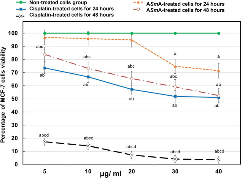
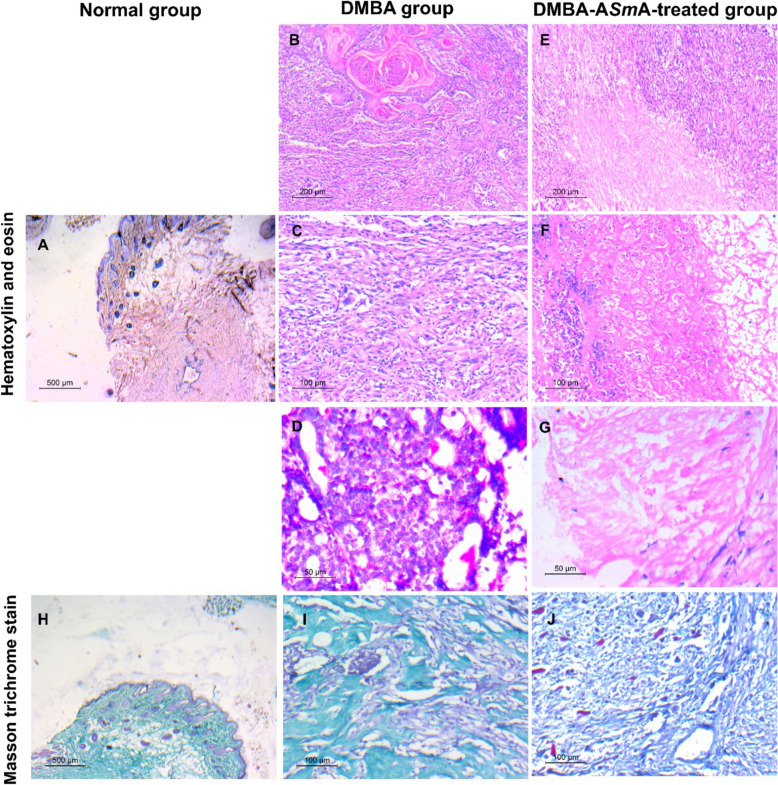
Results: In vitro, autoclaved Schistosoma mansoni antigen significantly reduced MCF-7 cell viability in a dose- and time-dependent manner. In vivo, autoclaved Schistosoma mansoni antigen treatment significantly reduced tumour weight and volume, improved liver enzyme levels, increased tumour necrosis, and decreased fibrosis. Immunohistochemical analysis revealed decreased Ki-67 and CD31 expression, indicating reduced cell proliferation and angiogenesis, respectively. Autoclaved Schistosoma mansoni antigen also enhanced immune responses by increasing CD8+ T cells infiltration and decreasing FoxP3+ expression, resulting in a higher CD8+ T cells/FoxP3+ ratio within the tumour microenvironment. Notably, PD-L1 expression was also downregulated, suggesting potential immune checkpoint inhibition.
Conclusions: Autoclaved Schistosoma mansoni antigen demonstrated potent anti-neoplastic activity, significantly reducing tumour growth and modulating the immune response within the tumour microenvironment. These results highlight autoclaved Schistosoma mansoni antigen's potential as a novel immunotherapy for breast cancer.
Keywords: Schistosoma; Breast cancer; MCF-7; Parasite immunotherapy.
© 2025. The Author(s).
Conflict of interest statement
Declarations. Ethics approval and consent to participate: All experimental animals were handled following the ARRIVE guidelines for animal care and in compliance with the Institutional Animal Care and Use Committee in the Faculty of Medicine, Alexandria University (IACUC; 0201715). Consent for publication: Not applicable. Competing interests: The authors declare no competing interests.
Figures


References
-
- World Health Organization. worldwide cancer statistics. Geneva: World Health Organization; 2024.
-
- Kaur R, Bhardwaj A, Gupta S. Cancer treatment therapies: traditional to modern approaches to combat cancers. Mol Biol Rep. 2023;50(11):9663–76. 10.1007/s11033-023-08809-3. - PubMed
-
- World Health Organization. Cancer Fact sheet. Geneva: World Health Organization; 2023.
MeSH terms
Substances
LinkOut - more resources
Full Text Sources
Medical
Research Materials
