. 2025 May-Jun;72(3):e70010.
doi: 10.1111/jeu.70010.

Novel Foraging Mechanisms in Atypical Excavate Flagellates

Affiliations

Novel Foraging Mechanisms in Atypical Excavate Flagellates

Sei Suzuki-Tellier et al. J Eukaryot Microbiol. 2025 May-Jun.

Abstract

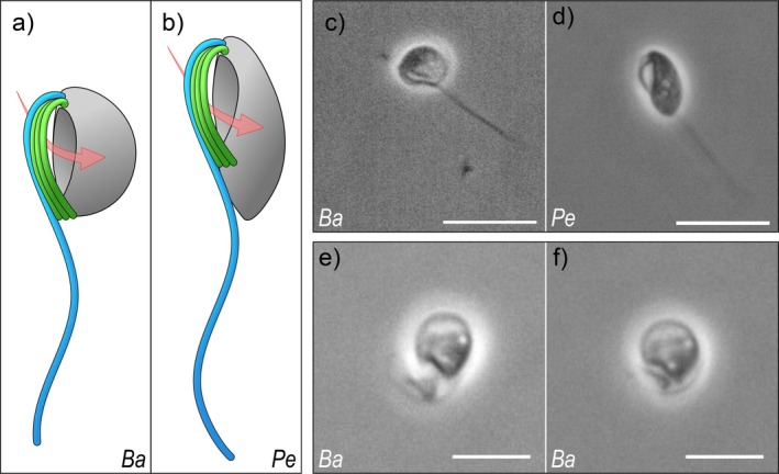
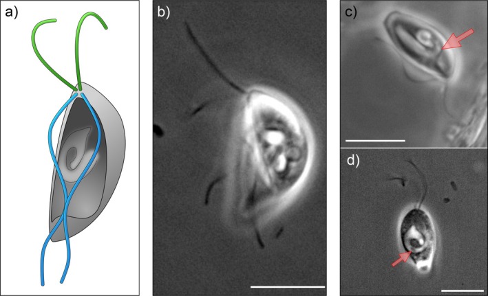
Most excavates, a paraphyletic assemblage of flagellates, typically present an active vaned flagellum that drives a feeding current through a ventral groove for predation. However, some have "atypical" morphologies. Here, we describe the foraging mechanisms in heteroloboseid flagellates (Discoba) that have a groove but lack the seemingly crucial vane. The percolomonads barbeliid AE-1 and Percolomonas doradorae form a functional vane with four adjacent flagella undulating with lateral asymmetry, creating an erratic flow that rapidly "sucks" water into the groove and expels it on the other side. This flow attenuates rapidly away from the cell, consistent with the flagellar pump acting as an instantaneous point sink. Conversely, Pharyngomonas kirbyi generates a continuous flow through the groove with two asynchronously moving posterior flagella. Despite the unexplained fluid dynamics, this flow has a further reach, consistent with describing the flagellar pump as a point force (stokeslet). While the volumetric flow rate through the groove-a measure of the maximum potential clearance rate-of the two percolomonads is similar to clearance rates estimated for other phagotrophic flagellates, it is an order of magnitude lower for Ph. kirbyi, which may afford lower rates due to high prey concentration in its hypersaline environment.

Keywords: clearance rate; feeding current; heteroloboseids; prey capture; vaneless flagella; ventral feeding groove.


Figures

FIGURE 1
FIGURE 1
Morphology and flagellar arrangement of barbeliid AE‐1 (Ba) and Percolomonas doradorae (Pe). Schematic representations (a, b) and phase‐contrast microscopy imaging (c–f) of barbeliid AE‐1 (a, c, and e, f) and Pe. doradorae (b and d). Red arrows (a and b) indicate the direction of the lateral feeding current. Images e and f show the ‘impermeable flagellar sheet’ that covers the width of the ventral groove along its entire length, from the perspective of the posterior end of the cell. Scale bars: 10 μm (c, d) and 5 μm (e, f).
FIGURE 2
FIGURE 2
Flow fields generated by barbeliid AE‐1 (a), Percolomonas doradorae (b), and Pharyngomonas kirbyi (c). For all species, the anterior end of the cell is at the top and the posterior end at the bottom of the panel. Barbeliid AE‐1 and Pe. doradorae are positioned laterally, and the yellow shadowing marks the area of the flagellar beat wave. Ph. kirbyi is viewed ventrally. The direction of the flow is represented chromatically: Originating from white/pale shades and moving toward intense colors. The yellow dots are the spatio‐temporal resolution of each particle tracking analysis, where time steps are 3.3 ms in panels (a) and (b), and 13.3 ms in panel (c).
FIGURE 3
FIGURE 3
Morphology and flagellar arrangement of Pharyngomonas kirbyi. Schematic representation (a) and phase‐contrast microscopy imaging (b–d): Panel a shows a ventral view of the cell, while panel b is a lateral perspective; (c) Ph. kirbyi is attached to the surface with an anterior flagellum with a visible entrance of the cytopharynx (red arrow) at the floor of the groove; (d) ventral view of the cytopharynx channel that spirals toward the ingestion site with a bacterium (red arrow) inside. Scale bars: 10 μm.

References

    1. Asadzadeh, S. S. , Walther J. H., Andersen A., and Kiørboe T.. 2022. “Hydrodynamic Interactions Are Key in Thrust‐Generation of Hairy Flagella.” Physical Review Fluids 7: 073101. 10.1103/PhysRevFluids.7.073101. - DOI
    1. Balmuth, W. , Bradbury P. C., and Schuster F. L.. 1983. “Ultrastructure of the Amoeboflagellate Tetramitus rostratus 1.” Journal of Protozoology 30: 445–455. 10.1111/j.1550-7408.1983.tb02946.x. - DOI - PubMed
    1. Boenigk, J. , and Arndt H.. 2000. “Particle Handling During Interception Feeding by Four Species of Heterotrophic Nanoflagellates.” Journal of Eukaryotic Microbiology 47: 350–358. 10.1111/j.1550-7408.2000.tb00060.x. - DOI - PubMed
    1. Brugerolle, G. , and Simpson A. G. B.. 2004. “The Flagellar Apparatus of Heteroloboseans.” Journal of Eukaryotic Microbiology 51: 96–107. 10.1111/j.1550-7408.2004.tb00169.x. - DOI - PubMed
    1. Carduck, S. , Nitsche F., Rybarski A., Hohlfeld M., and Arndt H.. 2021. “Diversity and Phylogeny of Percolomonads Based on Newly Discovered Species From Hypersaline and Marine Waters.” European Journal of Protistology 80: 125808. 10.1016/j.ejop.2021.125808. - DOI - PubMed

MeSH terms

LinkOut - more resources