. 2025 Dec;30(1):2491845.
doi: 10.1080/13510002.2025.2491845. Epub 2025 Apr 17.

Oxidation of human mitochondrial RNA strongly potentiates immunostimulation in an interferon-associated manner

Affiliations

Oxidation of human mitochondrial RNA strongly potentiates immunostimulation in an interferon-associated manner

Hung-Yun Lin et al. Redox Rep. 2025 Dec.

Abstract

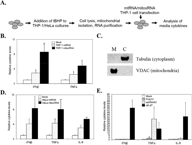
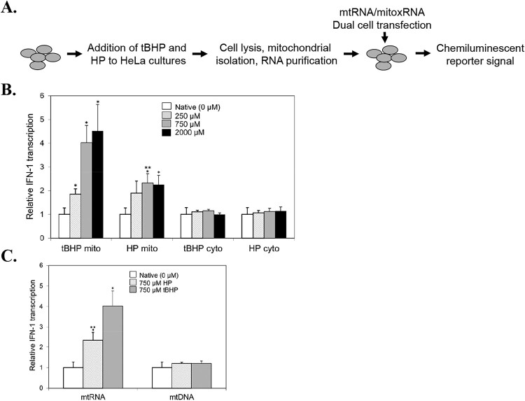
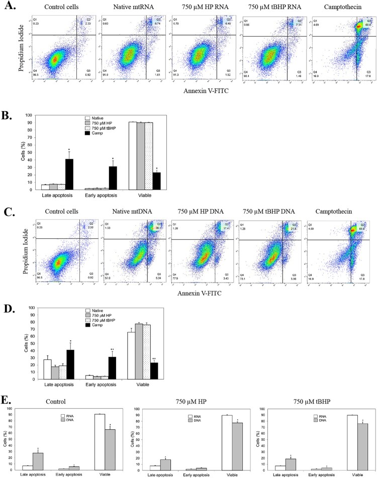
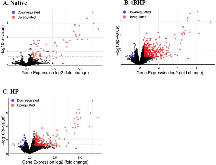
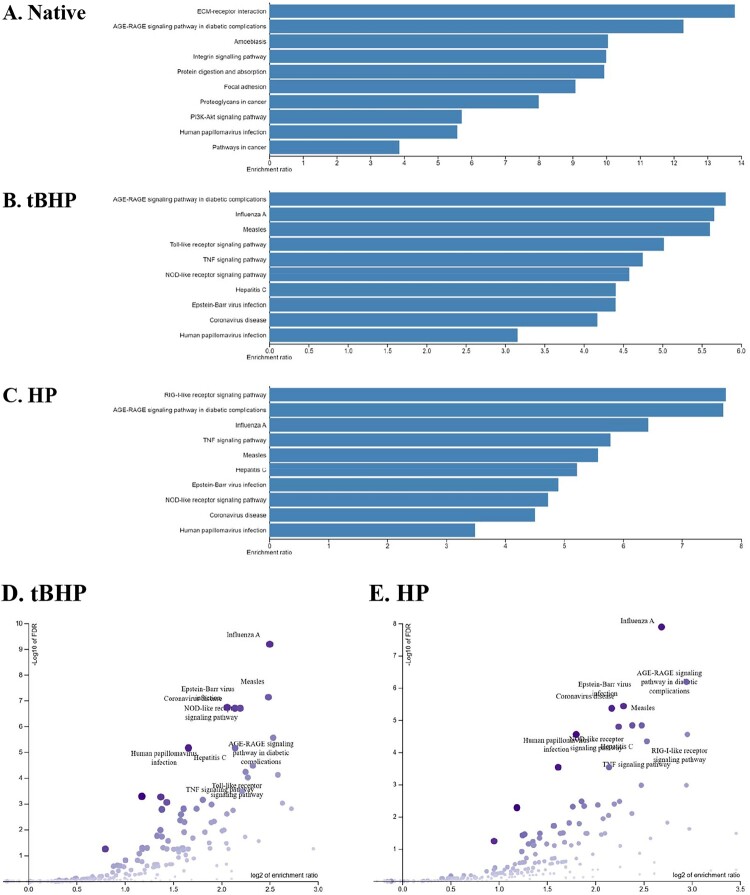
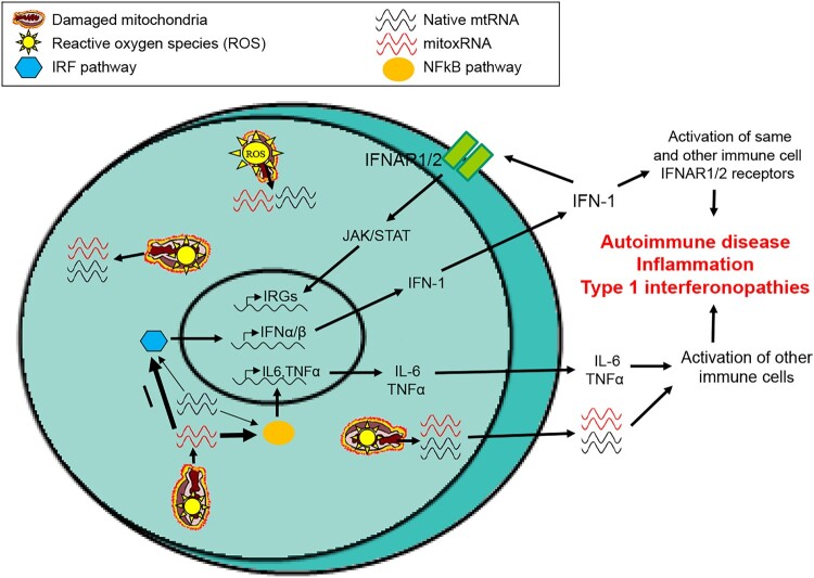
Inflammation is associated with a wide range of medical conditions, most leading causes of death, and high healthcare costs. It can thus benefit from new insights. Here we extended previous studies and found that oxidation of human native mtRNA to 'mitoxRNA' strongly potentiated IFNβ and TNFα immunostimulation in human cells, and that this newly identified type 1 interferon potentiation was transcriptional. This potentiation was significantly greater than with mtDNA oxidation, and t-butylhydroperoxide (tBHP) oxidation of RNA was more proinflammatory than hydrogen peroxide (HP). mtRNA triggered a modest increase in apoptosis that was not potentiated by oxidation, and mtDNA triggered a much greater increase. For native mtRNA, we found that chloroquine-inhibitable endosomes and MDA5 are key signaling pathways for IFNβ and TNFα production. For mitoxRNAs, RNAseq revealed a major increase in both tBHP- and HP-mitoxRNA modulated genes compared with native mtRNA. This increase was very prominent for interferon-related genes, identifying them as important mediators of this powerful oxidation effect. Moderately different gene modulations and KEGG pathways were observed for tBHP- versus HP-mitoxRNAs. These studies reveal the profound effect that mitochondrial RNA oxidation has on immunostimulation, providing new insights into DAMP inflammation and identifying potential therapeutic targets to minimize DAMP mtRNA/mitoxRNA-mediated inflammation.

Keywords: Mitochondria; T-butylhydroperoxide; human; hydrogen peroxide; immunostimulation; oxidative stress; proinflammatory cytokines; type 1 interferons.


Conflict of interest statement

No potential conflict of interest was reported by the author(s).

Figures

Figure 1.
Figure 1.
Immunostimulation of THP-1 cell IFNβ and proinflammatory cytokines with oxidized mtRNA. (a) General experimental strategy. (b) THP-1 cell native mtRNA and mitoxRNA induction of cytokines in THP-1 cells 20 h after transfection (N = 8). (c) Subcellular fraction analysis by Western immunoblotting using antibodies to the mitochondrial marker VDAC and the cytoplasmic marker alpha tubulin. M, mitochondrial fraction; C, cytoplasmic fraction. (d) HeLa cell native mtRNA and mitoxRNA induction of cytokines in THP-1 cells 20 h after transfection (N = 3–6). (e) Positive control ligand (polyI:C, ssRNA40, and dA:dT) induction of cytokines in THP-1 cells 20 h after transfection (N = 3–7). Mock transfection values were normalized to 1.0. Values represent means ± SEM. *, significant difference compared with Mock transfection using the Student’s t-test at p < .05. **, significant difference between native mtRNA and mitoxRNA.
Figure 2.
Figure 2.
Type 1 interferon (IFN-1) immunostimulation at the transcriptional level using Dual cells. (a) General experimental strategy. (b) Dual cell native mtRNA and mitoxRNA IFN-1 interferon transcription 20 h after transfection (N = 3–10). Mito, mitochondrial fraction; cyto, cytoplasmic fraction. (c) Comparison of Dual cell mitoxRNA versus mitoxDNA induction of IFN-1 transcription 20 h after transfection using 750 µM tBHP and 750 µM HP (N = 6–10). Mock transfection values were normalized to 1.0. Values represent means ± SEM. *, significant difference compared with Native using the Student’s t-test at p < .05. **, significant difference comparing 750 µM tBHP mitoxRNA with 750 µM HP mitoxRNA using the Student’s t-test at p < .05.
Figure 3.
Figure 3.
Effect of mtRNA and mitoxRNA on apoptosis of THP-1 cells. (a) Mitochondrial RNA-transfected cells were washed and resuspended and FITC Annexin and propidium iodide (PI) added, incubated, and analyzed by flow cytometry at 561 nm (PI) and 488 (FITC). The proapoptotic agent Camptothecin was used as a positive control. (b) Plotted data. (c) Mitochondrial DNA-transfected cells prepared and analyzed as above. (d) Plotted data for mitochondrial DNA. (e) Comparison of mtDNA and mitoxDNA effects on apoptosis. For (b, d) and (e) plotted data, the Y-axis ‘Cells (%)’ refers to each designated subpopulation as a percentage of total cells.
Figure 4.
Figure 4.
Comparison of mtDNA versus mitoxDNA forward and side-scatter. (a) Representative RNA analysis and (b) representative DNA analysis. Bottom panel shows Q2 and Q4 signals gated from both treatments and forward (FSC) and side-scatter (SSC) assessed.
Figure 5.
Figure 5.
MDA5 and endosome in native mtRNA immunostimulation. (a) THP-1 cells were pretreated with siRNA or CQ followed by HeLa mtRNA transfection and TNFα/IFNβ analysis. Data means (N = 6) ± SEM. *, significant difference vs Cont siRNA, Students t-test at p < .05. (b) MDA5 knockdown Western blot.
Figure 6.
Figure 6.
RNAseq heat maps. Hierarchal cluster heat maps for (a) Native, differentially expressed genes for mock- versus native mtRNA-transfected (76 modulated genes detected), (b) tBHP (TB), differentially expressed genes for mock- versus tBHP mitoxRNA-transfected (474 modulated genes), (c) HP, differentially expressed genes for mock- versus HP mitoxRNA-transfected (308 modulated genes). For above, (N = 3–4). Z-score scale bars based on Log2 FC are also shown. Blue, downregulated; red, upregulated.
Figure 7.
Figure 7.
RNAseq volcano plots. Volcano plots comparing samples described in Figure 6.
Figure 8.
Figure 8.
GOSlim summary. GOSlim summary of genes differentially altered in (a) native mtRNA-transfected (‘Native’) cells, and (b) tBHP mitoxRNA-transfected (‘tBHP’) cells. Bar charts for Biological Process (red), Cellular Component (blue), and Molecular Function (green) categories are presented with bar heights representative of the number of genes observed in the category.
Figure 9.
Figure 9.
ORA analyses of gene ontology biological processes. Bar graph representations of top KEGG pathway enrichment analysis results in order. (a) Native, mock- versus native mtRNA transfection, (b) tBHP, mock- versus tBHP mitoxRNA-transfected, and (c) HP, mock- versus HP mitoxRNA-transfected. All FDR ≤ 0.05. (d) Volcano plot of above ‘B’ tBHP, mock- versus tBHP mitoxRNA-transfected, and (e) Volcano plot of above ‘C’ HP, mock- versus tBHP mitoxRNA-transfected. For the volcano plots, −log10 of FDR (Y-axis) is plotted against the log2 enrichment ratio (X-axis) with dot size and color proportional to the size of the category and dot position to the category significance.
Figure 10.
Figure 10.
Model of mitochondrial RNA immunostimulation in stressed cell emphasizing type 1 interferon. Proposed model of native mtRNA and mitoxRNA release, signaling and pathological effects.

Similar articles

References

    1. Slavich GM. Understanding inflammation, its regulation, and relevance for health: a top scientific and public priority. Brain Behav Immun. 2015;45:13–14. doi:10.1016/j.bbi.2014.10.012 - DOI - PMC - PubMed
    1. Cifuentes M, Verdejo HE, Castro PF, et al. . Low-grade chronic inflammation: a shared mechanism for chronic diseases. Physiology (Bethesda). 2025;40(1):0. - PubMed
    1. Mathew A, Lindsley TA, Sheridan A, et al. . Degraded mitochondrial DNA is a newly identified subtype of the damage associated molecular pattern (DAMP) family and possible trigger of neurodegeneration. J Alzheimers Dis. 2012;30:617–627. doi:10.3233/JAD-2012-120145 - DOI - PubMed
    1. Saxena AR, Gao LY, Srivatsa S, et al. . Oxidized and degraded mitochondrial polynucleotides (DeMPs), especially RNA, are potent immunogenic regulators in primary mouse macrophages. Free Radic Biol Med. 2017;104:371–379. doi:10.1016/j.freeradbiomed.2017.02.009 - DOI - PubMed
    1. Ilic Z, Saxena AR, Periasamy S, et al. . Control (native) and oxidized (DeMP) mitochondrial RNA are proinflammatory regulators in human. Free Radic Biol Med. 2019;143:62–69. doi:10.1016/j.freeradbiomed.2019.07.019 - DOI - PubMed

LinkOut - more resources