. 2025 Jun 1;328(6):L757-L771.
doi: 10.1152/ajplung.00380.2024. Epub 2025 Apr 17.

Protocol for differentiating primary human small airway epithelial cells at the air-liquid interface

Affiliations

Protocol for differentiating primary human small airway epithelial cells at the air-liquid interface

Yu Par Aung Myo et al. Am J Physiol Lung Cell Mol Physiol. .

Abstract

The air-liquid interface (ALI) culture is an important tool in pulmonary research as it models the physiological lung where the epithelium is apically exposed to air and basally to the endothelium and interstitium. Although there is an abundance of research that uses primary human bronchial epithelial cells (HBECs) to study larger airways, small airway epithelial cells (SAECs) are an untapped resource in comparison. Primary SAECs are a valuable cell population as they enable the study of pathologies in the bronchioles and are also a favorable surrogate for primary alveolar epithelial cells, which are invasive to collect from patients. Currently, there are limited resources on how to culture and differentiate SAECs at the ALI. Here, we provide an optimized, detailed protocol to address this knowledge gap. Key culture conditions that determine the quality and uniformity of differentiated SAECs include cell passage number, pH changes caused by media exhaustion and incubator CO2, seeding density, and collagen coating of the expansion flask and inserts. We also describe a FITC-dextran permeability assay to measure SAEC barrier integrity both as a pretest to select uniform wells with strong barrier integrity before an experiment and as a post-test to evaluate treatment effects afterward. The utility of the differentiated SAEC ALI model to ask biologically relevant questions is demonstrated by increased cytokine (IL-8, MIF, and CXCL-10) production and/or epithelial damage following exposure to cigarette smoke, lipopolysaccharide (LPS) or poly(I:C).NEW & NOTEWORTHY SAECs are not commonly used in pulmonary research, and this is reflected in a lack of literature on both SAEC primary research and methodological reports. Primary SAECs are an important resource as they enable the study of the small airways, which are implicated in a variety of pulmonary diseases, including chronic obstructive pulmonary disease (COPD). The detailed protocol described here bridges the knowledge gap on how to successfully differentiate primary human SAECs at the ALI.

Keywords: air-liquid interface; differentiation; methods; primary human cells; small airway epithelial cells.


Conflict of interest statement

DISCLOSURES

No conflicts of interest to disclose.

Figures
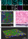
Figure 1.
Figure 1.. SAECs differentiate into a complex one-to-two cell layer composed of ciliated, club and basal cells.
SAECs were differentiated in Pneumacult ALI-S media for 4 weeks after air-lift at the ALI. (A) Immunofluorescence staining of acetylated tubulin (AcTub, marker for ciliated cells), CC10 (marker for club cells), keratin5 (Krt5, marker for basal cells), E-cadherin (E-cad, adherens junction protein), zonula occludens-1 (ZO-1, tight junction protein) and nuclei (DAPI). Scale bars = 50 μm. (B) Top-down and cross-section view of an immunofluorescence confocal z-stack. Green = AcTub, red = CC10, white = Krt5, yellow = ZO-1, blue = DAPI. (C) Top-down and cross-section H&E stain. Top-down view appears freckled because the pores in the insert membrane absorb some dye. (D) 3D rendering of an immunofluorescence confocal z-stack. Colors for target proteins are the same as (B). Cultures shown are from 2 different donors (A = donors 2 and 3, B, C and D = donor 3). Images were acquired using the Zeiss LSM700 (A), Leica STELLARIS (B, D) and Zeiss Axio Imager.Z2 (C) microscopes. Representative images are shown. A video file of the 3D rendering of the ALI cell culture is available as an .mp4 file downloadable at doi:10.6084/m9.figshare.c.7683176
Figure 2.
Figure 2.. Protocol C produced differentiated SAECs with high barrier integrity.
(A) Schematic of the FITC-dextran permeability assay created with BioRender.com. (B) SAECs with 30% and 50% confluency were incubated with FITC-dextran for 0, 30, 60 and 120 minutes (n = 1 – 3). (C) The barrier integrity of differentiated donor 1 SAECs from protocols A, B and C was measured by incubating with FITC-dextran for 120 minutes (n = 71 – 72). (D) Comparison of protocol C across 4 different donors to assess reproducibility. Differentiated SAEC cultures were incubated with FITC-dextran for 120 minutes (n = 48 – 71). (E) Comparison of FITC-dextran leak and TEER at baseline for donor 3 SAECs (n = 7). Two-way ANOVA with post-hoc Tukey was performed for (B) and one-way ANOVA with post-hoc Tukey was performed for (C) and (D).
Figure 3.
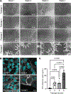
Figure 3.. Passage number is a crucial factor in the formation of a contiguous epithelial barrier.
(A) Representative images of passage 3, 4, 5 and 6 donor 1 SAECs taken over 4 weeks of differentiation after airlift in Pneumacult ALI-S media under protocol C. White arrows outline gaps in the barrier. Scale bars = 600 μm. (B) Donor 1 SAECs were immunostained for E-cadherin (white) and nuclei (DAPI, cyan) at the end of 4 weeks of differentiation. (C) Comparison of barrier epithelial integrity between passage 3, 4, 5 and 6 donor 1 SAECs at the end of week 4 using the FITC-dextran permeability assay (120-minute incubation). Images were taken with the EVOS M7000 (A) and Zeiss LSM700 (B) microscopes. Representative images are shown. One-way ANOVA with post-hoc Tukey was performed for statistics. n= 3 – 5.
Figure 4.
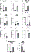
Figure 4.. Cigarette smoke causes epithelial damage and induces pro-inflammatory cytokine production.
SAECs were differentiated at the ALI following protocol C and exposed to cigarette smoke (CS) for 4 consecutive days (30 minutes each day). On day 5, FITC-dextran permeability assays (A, B, C, J) were performed, basal media was collected for IL-8 (D, E, F) and MIF (G, H, I) ELISAs and TEER was measured (J). Data shown are from 3 different donors (donor 1 = A, D, G donor 2 = B, E, H, donor 3 = C, F, I, J) Unpaired, two-tailed student’s t-tests were run for statistics. n = 4 – 7 replicates per donor.
Figure 5.
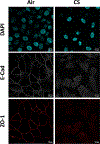
Figure 5.. Cigarette smoke decreases both E-cadherin and zonula occludens-1.
Donor 3 SAECs were differentiated at the ALI following protocol C and exposed to cigarette smoke (CS) for 4 consecutive days (30 minutes each day). On day 5, inserts were fixed for E-cadherin (white), ZO-1 (red) and nuclei (DAPI, cyan) immunofluorescence staining. Images were taken with the Zeiss LSM700 microscope. Representative images are shown.
Figure 6.
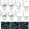
Figure 6.. Poly(I:C) and Pseudomonas aeuroginosa LPS induce cytokine production but not epithelial damage.
Donor 4 SAECs were differentiated under protocol C and stimulated with 0.1 – 2.5 μg/mL high molecular weight poly(I:C) (A-D) and 10 – 1000 ng/mL Pseudomonas aeuroginosa LPS (E-H). 24h after stimulation, basal media was collected for IL-8 (A, E), MIF (B, F) and CXCL-10 (C, G) ELISAs. FITC-dextran permeability assays (D, H) were performed. (I) SAECs were immunostained for E-cadherin (white) and nuclei (DAPI, cyan). Images were taken with the Zeiss LSM700 microscope. One-way ANOVA with post-hoc Dunnett (A, B, C, E, F, G) and Tukey (D, H) was performed for statistics. n=3.

References

    1. Davis JD, Wypych TP. Cellular and functional heterogeneity of the airway epithelium. Mucosal Immunol 14: 978–990, 2021. doi: 10.1038/s41385-020-00370-7. - DOI - PMC - PubMed
    1. Doni Jayavelu N, Altman MC, Benson B, Dufort MJ, Vanderwall ER, Rich LM, White MP, Becker PM, Togias A, Jackson DJ, Debley JS. Type 2 inflammation reduces SARS-CoV-2 replication in the airway epithelium in allergic asthma through functional alteration of ciliated epithelial cells. J Allergy Clin Immunol 152: 56–67, 2023. doi: 10.1016/J.JACI.2023.03.021. - DOI - PMC - PubMed
    1. Bulcaen M, Kortleven P, Liu RB, Maule G, Dreano E, Kelly M, Ensinck MM, Thierie S, Smits M, Ciciani M, Hatton A, Chevalier B, Ramalho AS, Casadevall i Solvas X, Debyser Z, Vermeulen F, Gijsbers R, Sermet-Gaudelus I, Cereseto A, Carlon MS. Prime editing functionally corrects cystic fibrosis-causing CFTR mutations in human organoids and airway epithelial cells. Cell Rep Med 5: 101544, 2024. doi: 10.1016/J.XCRM.2024.101544. - DOI - PMC - PubMed
    1. Wasnick R, Korfei M, Piskulak K, Henneke I, Wilhelm J, Mahavadi P, Dartsch RC, von der Beck D, Koch M, Shalashova I, Weiss A, Klymenko O, Askevold I, Fink L, Witt H, Hackstein H, El Agha E, Bellusci S, Klepetko W, Königshoff M, Eickelberg O, Schermuly RT, Braun T, Seeger W, Ruppert C, Guenther A. Notch1 Induces Defective Epithelial Surfactant Processing and Pulmonary Fibrosis. Am J Respir Crit Care Med 207: 283–299, 2023. doi: 10.1164/RCCM.202105-1284OC. - DOI - PubMed
    1. McCarthy CE, Duffney PF, Gelein R, Thatcher TH, Elder A, Phipps RP, Sime PJ. Dung biomass smoke activates inflammatory signaling pathways in human small airway epithelial cells. Am J Physiol Lung Cell Mol Physiol 311: L1222–L1233, 2016. doi: 10.1152/ajplung.00183.2016. - DOI - PMC - PubMed

LinkOut - more resources