

In vitro wound healing effects of postbiotics derived from the gut microbiota of long-lived blind mole rats, a model of healthy ageing
- PMID: 40247718
- PMCID: PMC12006833
- DOI: 10.1111/wrr.70023
In vitro wound healing effects of postbiotics derived from the gut microbiota of long-lived blind mole rats, a model of healthy ageing
Abstract
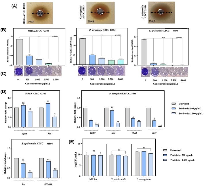
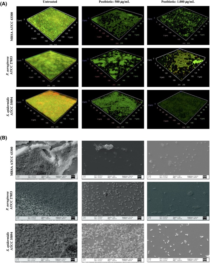
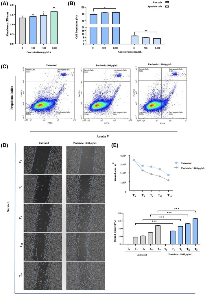
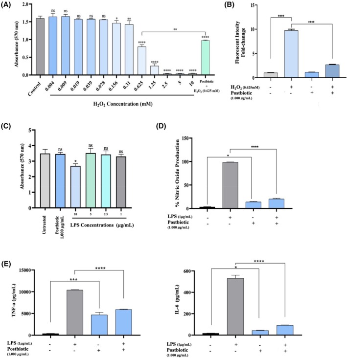
Chronic wounds represent a global public health burden to patients and healthcare professionals worldwide. Considering the unmet need for safe and effective therapeutic approaches for wound healing, research on discovering new bioactive materials that support all stages of wound healing is gaining importance. In this study, the wound-healing activity of postbiotics obtained from Limosilactobacillus reuteri EIR/Spx-2, isolated from the gut microbiota of long-lived blind mole rats (Nannospalax xanthodon), was investigated. Our results demonstrated that postbiotics exhibited a strong inhibitory effect against important skin pathogens, eliminated their biofilm formation, and downregulated the expression of genes involved in their quorum-sensing regulatory mechanisms. Furthermore, treatment with postbiotics resulted in a significant increase (23.82% ± 2.11%) in L929 fibroblast cell proliferation. Additionally, postbiotics applied on scratched fibroblast monolayer significantly accelerated the re-epithelialization by 66.78% ± 3.74%. The treatment also increased the mRNA expression and protein levels of COL1A1 in the early healing phase. Moreover, the intracellular ROS levels of L929 cells suppressed by H2O2 were significantly reduced, which could be attributed to the content of flavonoids (4.8 mg/g) and phenolic compounds (7.12 mg/g) in postbiotics, as well as their DPPH scavenging activity. After treatment with postbiotics, the mRNA levels of IL-6 (5.77-fold) and TNF-α (1.76-fold) and the amount of NO (79.25% ± 3.18%) were significantly decreased in LPS-induced murine macrophages. The diverse metabolite profile of postbiotics, as characterised using chromatographic techniques, exhibited a strong correlation with their biological activity across all stages of the wound healing process, highlighting their potential as promising candidates for wound healing applications.
Keywords: biofilms; infection; inflammation Limosilactobacillus reuteri; postbiotics; proliferation; wound healing.
© 2025 The Author(s). Wound Repair and Regeneration published by Wiley Periodicals LLC on behalf of The Wound Healing Society.
Conflict of interest statement
Authors declare no conflicts of interest.
Figures

Similar articles
-
Lafoensia pacari A. St.-Hil.: Wound healing activity and mechanism of action of standardized hydroethanolic leaves extract.J Ethnopharmacol. 2018 Jun 12;219:337-350. doi: 10.1016/j.jep.2018.02.038. Epub 2018 Mar 6. J Ethnopharmacol. 2018. PMID: 29501673
-
A Novel Effective Formulation of Bioactive Compounds for Wound Healing: Preparation, In Vivo Characterization, and Comparison of Various Postbiotics Cold Creams in a Rat Model.Evid Based Complement Alternat Med. 2021 Dec 7;2021:8577116. doi: 10.1155/2021/8577116. eCollection 2021. Evid Based Complement Alternat Med. 2021. PMID: 34917159 Free PMC article.
-
Oat-based postbiotics ameliorate high-sucrose induced liver injury and colitis susceptibility by modulating fatty acids metabolism and gut microbiota.J Nutr Biochem. 2024 Mar;125:109553. doi: 10.1016/j.jnutbio.2023.109553. Epub 2023 Dec 24. J Nutr Biochem. 2024. PMID: 38147914
-
Postbiotics-A Step Beyond Pre- and Probiotics.Nutrients. 2020 Jul 23;12(8):2189. doi: 10.3390/nu12082189. Nutrients. 2020. PMID: 32717965 Free PMC article. Review.
-
Postbiotics-peptidoglycan, lipoteichoic acid, exopolysaccharides, surface layer protein and pili proteins-Structure, activity in wounds and their delivery systems.Int J Biol Macromol. 2024 Aug;274(Pt 1):133195. doi: 10.1016/j.ijbiomac.2024.133195. Epub 2024 Jun 15. Int J Biol Macromol. 2024. PMID: 38885869 Review.
References
-
- Martins‐Green M. Cutaneous chronic wounds: a worldwide silent epidemic. Open Access Government. 2023;38(1):51‐53.
-
- World Health Organization . 2022. https://www.who.int/news-room/fact-sheets/detail/ageing-and-health.
MeSH terms
LinkOut - more resources
Full Text Sources
Miscellaneous
