

Extra axial medulloblastoma of the cerebellopontine angle: A rare case report
- PMID: 40247957
- PMCID: PMC12005853
- DOI: 10.1016/j.radcr.2025.03.034
Extra axial medulloblastoma of the cerebellopontine angle: A rare case report
Abstract
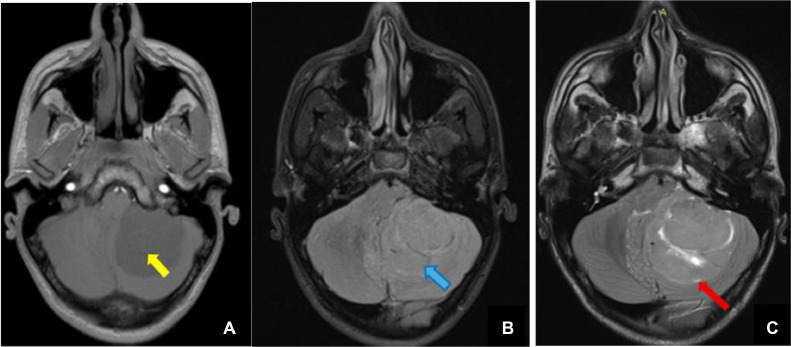
Medulloblastoma is the most frequent malignant brain tumor in children. Originating in the cerebellum, they are typically intra-axial tumors. In adults, they represent less than 1% of brain tumors. However, the occurrence of extra-axial medulloblastoma is possible but extremely rare, and slightly more frequent in the adult population. We present a rare case of extra-axial medulloblastoma, diagnosed in a 22-year-old male, through advanced imaging techniques, followed by confirmation through anatomopathological examination. This case calls attention to the necessity of knowledge of the various diagnostic possibilities when interpreting radiological images, leading to enhanced patient care and furthering our understanding of these exceptional entities.
Keywords: Cerebello-pontine angle; Extra axial; Histopathology; MRI; Medulloblastoma; Schwannoma.
© 2025 The Authors. Published by Elsevier Inc. on behalf of University of Washington.
Figures


Similar articles
-
Extra-axial medulloblastoma in cerebello-pontine angle: A report of a rare case with literature review.Med J Islam Repub Iran. 2014 Jul 13;28:57. eCollection 2014. Med J Islam Repub Iran. 2014. PMID: 25405123 Free PMC article.
-
Extra-Axial Cerebello-Pontine Angle Medulloblastoma in an Infant: A Rare Case Report with Review of Literature.Asian J Neurosurg. 2021 Sep 14;16(3):447-451. doi: 10.4103/ajns.AJNS_79_21. eCollection 2021 Jul-Sep. Asian J Neurosurg. 2021. PMID: 34660353 Free PMC article. Review.
-
Extra-axial medulloblastoma in the cerebellopontine angle: Report of a rare entity with review of literature.J Pediatr Neurosci. 2016 Oct-Dec;11(4):331-334. doi: 10.4103/1817-1745.199477. J Pediatr Neurosci. 2016. PMID: 28217158 Free PMC article.
-
Extra-axial adult cerebellopontine angle medulloblastoma: Revisiting a rare entity.J Cancer Res Ther. 2022 Apr-Jun;18(3):770-773. doi: 10.4103/jcrt.JCRT_675_20. J Cancer Res Ther. 2022. PMID: 35900553
-
Posterior fossa extra-axial variations of medulloblastoma: a pictorial review as a primer for radiologists.Insights Imaging. 2021 Apr 6;12(1):43. doi: 10.1186/s13244-021-00981-z. Insights Imaging. 2021. PMID: 33822292 Free PMC article. Review.
References
-
- Mahapatra S., Amsbaugh M.J. StatPearls. StatPearls Publishing; Treasure IslandFL: 2023. Medulloblastoma.
-
- Tumeurs cérébrales . J. Philippon; Masson, Paris: 2004. du diagnostic au traitement; p. 285. ISBN 2-294-01288-7.
-
- Menon G., Krishnakumar K., Nair S. Adult medulloblastoma: clinical profile and treatment results of 18 patients. J Clin Neurosci. 2008;15:122–126. - PubMed
-
- Akay K.M., Erdogan E., Izci Y., Kaya A., Timurkaynak E. Medulloblastoma of the cerebellopontine angle. Neurol Med Chir. 2003;43:555–558. - PubMed
Publication types
LinkOut - more resources
Full Text Sources
