. 2025 Apr 16;27(Suppl 3):iii40-iii45.
doi: 10.1093/eurheartjsupp/suaf014. eCollection 2025 Mar.

When the conduction disturbance expresses a cardiomyopathy

Affiliations

When the conduction disturbance expresses a cardiomyopathy

Leonardo Calò et al. Eur Heart J Suppl. .

Abstract

Electrocardiogram may play a crucial role in the diagnostic workup of different cardiomyopathies. Electrocardiogram abnormalities in impulse generation and transmission may be an early marker of these insidious pathologies. Some findings are suggestive of definite disorders, and other findings are less sensitive and/or specific, but may orient towards a specific diagnosis in a patient with a peculiar phenotype. Electrocardiogram findings not only could help to early recognize affected patients but may also have an important prognostic role in evaluating disease evolution over time and identifying patients at higher risk of sudden cardiac death. Electrocardiogram reading and the careful interpretation of its features remain a cornerstone for orienting the diagnosis towards specific forms and provide useful tools for risk stratification.

Keywords: Arrhythmogenic cardiomyopathy; Cardiomyopathy; Conduction disturbances; Electrocardiogram.


Conflict of interest statement

Conflict of interest: none declared.

Figures
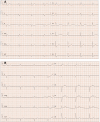
Figure 1
Figure 1
First-degree atrioventricular block and intra-ventricular conduction disturbances in patients with non-ischaemic cardiomyopathy. (A) The electrocardiogram of a 64-year-old man with arrhythmogenic left ventricular cardiomyopathy [carrier of a probable pathogenic variant in the Lamin A/C gene (c.949G>A, p.Glu317Lys)]. His electrocardiogram shows sinus bradycardia, first-degree atrioventricular block (PR 334 ms), left anterior fascicular block, and right bundle branch block. (B) The electrocardiogram of a 75-year-old man admitted to the hospital for dyspnoea and fatigue. His electrocardiogram shows extreme sinus bradycardia, first-degree atrioventricular block (PR 320 ms), incomplete right bundle branch block, and lack of q-wave in the left pre-cordial Leads LV, V6. A pseudo-infarct pattern was also present in Leads D3 and aVF and low QRS voltages in V1 and V2. During hospitalization, coronary arteries free of significant stenosis were found on coronary angiography, and symmetric left ventricular hypertrophy was found on echocardiogram. Subsequently, c-99m-3,3-diphosphono-1,2-propanedicarboxylic acid bone scan showed Grade 3 uptake of the tracer in the cardiac region. This finding, together with negative genetic analysis, supported the diagnosis of wild-type transthyretin cardiac amyloidosis.
Figure 2
Figure 2
Left anterior haemiblocks in arrhythmogenic and dilated cardiomyopathies. (A) Electrocardiogram of a 63-year-old man with dilated cardiomyopathy and severe left ventricular dysfunction. His electrocardiogram shows sinus rhythm with left axis deviation (QRS axis 68°, arrow). Note the presence of necrosis (QS pattern) in the inferior leads (D2, D3, and aVF) that simulate a left anterior haemiblock. (B) The absence of r in the inferior leads suggests extensive necrosis as demonstrated by the presence of inferior transmural late gadolinium enhancement on cardiac magnetic resonance imaging. (C) Electrocardiogram of a 37-year-old man with left arrhythmogenic cardiomyopathy, carrier of a probably pathogenic mutation affecting the DSP gene. His electrocardiogram shows, in addition to the presence of low peripheral voltages, the presence of a left anterior haemiblock (QRS axis ∼41°). The presence of a small r wave in the inferior location could hide the presence of fibrosis. The patient’s cardiac magnetic resonance (D) shows the presence of inferior sub-epicardial late gadolinium enhancement, however made suspicious by the presence of low peripheral voltages and the presence of a late depolarization current.
Figure 3
Figure 3
Left posterior haemiblock in arrhythmogenic cardiomyopathies. (A) Case report of a 21-year-old female admitted to the hospital due to an aborted cardiac arrest, which occurred at rest. The 12-lead surface electrocardiogram showed diffuse low QRS voltages, negative T waves in D3, and left posterior fascicular block. The electrocardiogram also showed polymorphic premature ventricular complexes characterized by right bundle branch block morphology, with a superior axis in the frontal plane, and left bundle branch block morphology, with an inferior axis morphology. The echocardiogram showed a mild depression of the left ventricular ejection fraction, estimated at 50%. Coronary angiography showed an accessory septal branch of the right coronary artery. Cardiac magnetic resonance imaging showed an area of late gadolinium enhancement in the sub-epicardial layer of the lateral wall of the left ventricle. (B) The electrocardiogram of a patient with arrhythmogenic cardiomyopathy with biventricular involvement reveals an extreme right axis deviation (A anomalous QRS +120°), with prominent anterior forces (S-wave depth in V1 <5 mm, R wave of V2 >15 mm, R/S ratio in V1 and V2 >2, rising R-wave from V1 to V3 and falling from V5 to V6, and lack of q-wave in left pre-cordial Leads LV, V6). These features suggest the possible concomitant presence of the left septal fascicular block together with the left posterior fascicular block. Diffuse low QRS voltages are present, especially in the peripheral leads. (C and D) Cardiac magnetic resonance images show late sub-endocardial gadolinium enhancement involving the right ventricle, septum, and inferior wall of the left ventricle and sub-epicardial late gadolinium enhancement of the lateral wall of the left ventricle (arrows).

References

    1. Elizari MV. The normal variants in the left bundle branch system. J Electrocardiol 2017;50:389–399. - PubMed
    1. Finocchiaro G, Merlo M, Sheikh N, De Angelis G, Papadakis M, Olivotto I et al. The electrocardiogram in the diagnosis and management of patients with dilated cardiomyopathy. Eur J Heart Fail 2020;22:1097–1107. - PubMed
    1. Crescenzi C, Silvetti E, Romeo F, Martino A, Bressi E, Panattoni G et al. The ECG in non-ischemic dilated cardiomyopathy. Eur Heart J Suppl 2023;25:C179–C184. - PMC - PubMed
    1. Silvetti E, Lanza O, Romeo F, Martino A, Fedele E, Lanzillo C et al. The pivotal role of ECG in cardiomyopathy. Front Cardiovasc Med 2023;10:1178163. - PMC - PubMed
    1. van Rijsingen IA, Arbustini E, Elliott PM, Mogensen J, Hermans-van Ast JF, van der Kooi AJ et al. Risk factors for malignant ventricular arrhythmias in lamin a/c mutation carriers: a European cohort study. J Am Coll Cardiol 2012;59:493–500. - PubMed

LinkOut - more resources