

Imaging the ex-vivo human cochlea using 1.3- μ m and 1.7- μ m optical coherence tomography
- PMID: 40248314
- PMCID: PMC12005953
- DOI: 10.1117/1.JBO.30.4.046007
Imaging the ex-vivo human cochlea using 1.3- μ m and 1.7- μ m optical coherence tomography
Abstract
Significance: There is no clinical imaging method to visualize the soft tissues of the human cochlea, which are crucial for sound transduction and are damaged in sensorineural hearing loss. Although optical coherence tomography (OCT) has been effective in small animal models, we show for the first time that it can image through the full thickness of the ex-vivo human otic capsule and resolve cochlear microstructures despite increased scattering.
Aim: We aim to investigate whether OCT could image the cochlea through the otic capsule. We compared 1.7 and OCT to test if the reduced scattering at provided any appreciable advantage for imaging the cochleae.
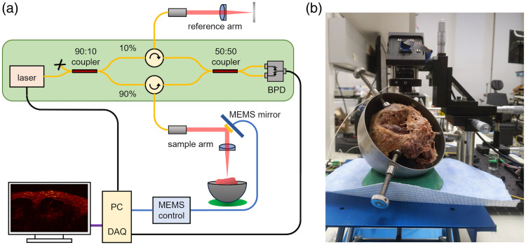
Approach: OCT interferometers were built for both 1.3 and wavelengths, using identical sample and reference arm optics in both systems. Imaging was performed on two fixed human temporal bones with intact cochleae. The interferometers were designed to allow seamless switching between 1.3 and OCT without disrupting the temporal bone during imaging.
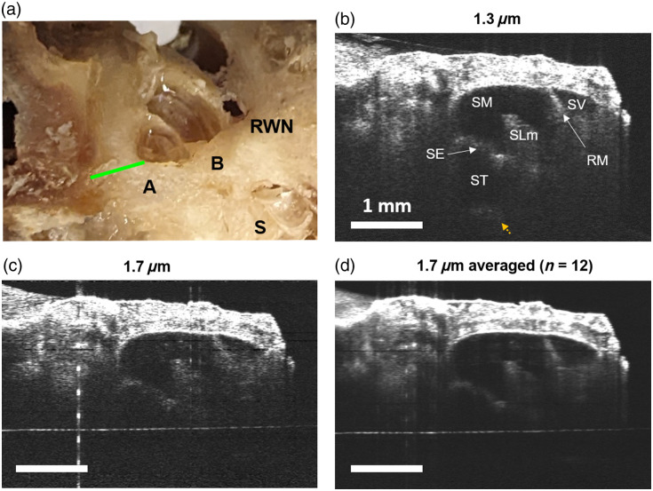
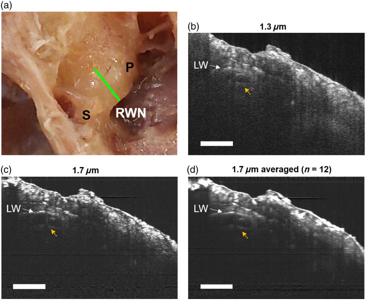
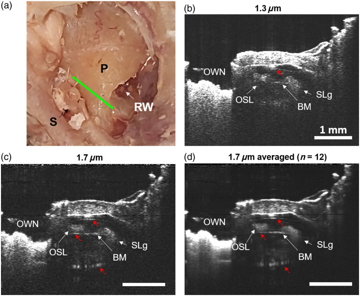
Results: We took volumetric OCT images at the base, apex, and hook regions of fixed ex-vivo human cochleae and compared the images taken at with those taken at . At both wavelengths, we could see through the otic capsule and identify cochlear structures. In some cases, OCT resulted in clearer images of the lateral wall, interior scala, and fine cochlear structures due to reduced multiple scattering at depth compared with .
Conclusions: We conclude that both and OCT can image through the human otic capsule, offering the potential for direct measurement of cochlear vibrometry or blood flow in living humans. Using light, we observed reduced multiple scattering in the otic capsule, leading to enhanced contrast of cochlear structures compared with . However, these improvements were marginal and came with trade-offs.
Keywords: cochleae; inner ear; optical coherence tomography; otolaryngology; otology.
© 2025 The Authors.
Figures
Similar articles
-
Optical coherence tomography (OCT) for detection of macular oedema in patients with diabetic retinopathy.Cochrane Database Syst Rev. 2015 Jan 7;1(1):CD008081. doi: 10.1002/14651858.CD008081.pub3. Cochrane Database Syst Rev. 2015. PMID: 25564068 Free PMC article.
-
Optical coherence tomography (OCT) for detection of macular oedema in patients with diabetic retinopathy.Cochrane Database Syst Rev. 2011 Jul 6;(7):CD008081. doi: 10.1002/14651858.CD008081.pub2. Cochrane Database Syst Rev. 2011. Update in: Cochrane Database Syst Rev. 2015 Jan 07;1:CD008081. doi: 10.1002/14651858.CD008081.pub3. PMID: 21735421 Updated.
-
Artificial intelligence for detecting keratoconus.Cochrane Database Syst Rev. 2023 Nov 15;11(11):CD014911. doi: 10.1002/14651858.CD014911.pub2. Cochrane Database Syst Rev. 2023. PMID: 37965960 Free PMC article.
-
Topical antiseptics for chronic suppurative otitis media.Cochrane Database Syst Rev. 2025 Jun 9;6(6):CD013055. doi: 10.1002/14651858.CD013055.pub3. Cochrane Database Syst Rev. 2025. PMID: 40484403
-
The Black Book of Psychotropic Dosing and Monitoring.Psychopharmacol Bull. 2024 Jul 8;54(3):8-59. Psychopharmacol Bull. 2024. PMID: 38993656 Free PMC article. Review.
References
Publication types
MeSH terms
Grants and funding
LinkOut - more resources
Full Text Sources
Miscellaneous
