Review
. 2025 Apr 3:15:1573373.
doi: 10.3389/fcimb.2025.1573373. eCollection 2025.

Phospholipid scramblase 1: a frontline defense against viral infections

Affiliations
Review

Phospholipid scramblase 1: a frontline defense against viral infections

Alina X Yang et al. Front Cell Infect Microbiol. .

Abstract

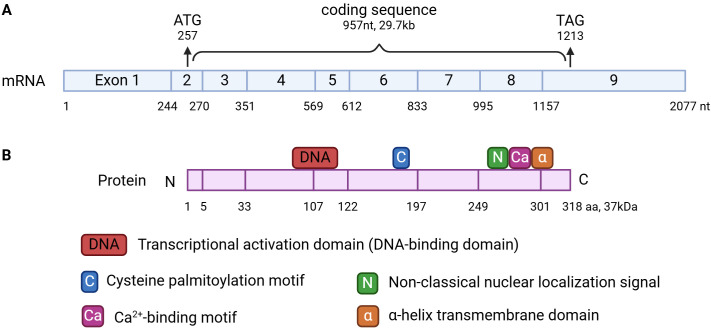
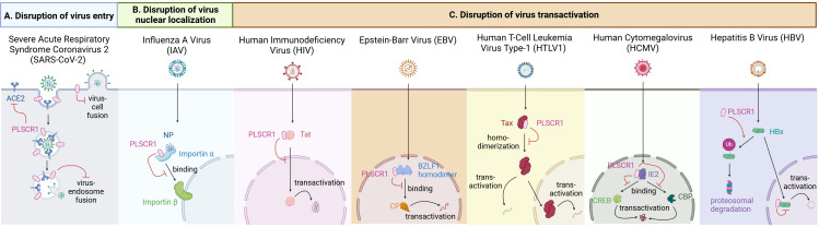
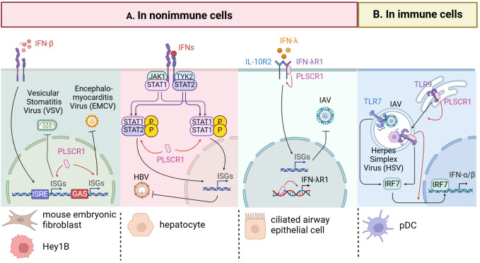
Phospholipid scramblase 1 (PLSCR1) is the most studied member of the phospholipid scramblase protein family. Its main function is to catalyze calcium (Ca2+)-dependent, ATP-independent, bidirectional and non-specific translocation of phospholipids between inner and outer leaflets of plasma membrane. Additionally, PLSCR1 is identified as an interferon-stimulated gene (ISG) with antiviral activities, and its expression can be highly induced by all types of interferons in various viral infections. Indeed, numerous studies have reported the direct antiviral activities of PLSCR1 through interrupting the replication processes of a variety of viruses, including entry of severe acute respiratory syndrome coronavirus 2 (SARS-CoV-2), nuclear localization of influenza A virus (IAV), and transactivation of human immunodeficiency virus (HIV), Epstein-Barr virus (EBV), human T-cell leukemia virus type-1 (HTLV1), human cytomegalovirus (HCMV) and hepatitis B virus (HBV). In addition to these direct antiviral activities, PLSCR1 also regulates endogenous immune components to defend against viruses in both nonimmune and immune cells. Such activities include potentiation of ISG transcription, activation of JAK/STAT pathway, upregulation of type 3 interferon receptor (IFN-λR1) and recruitment of Toll-like receptor 9 (TLR9). This review aims to summarize the current understanding of PLSCR1's multiple roles as a frontline defense against viral infections.

Keywords: Epstein-Barr virus; HBV - hepatitis B virus; HCMV (human cytomegalovirus); HIV; PLSCR1; SARS-CoV-2; antiviral; influenza A virus.


Conflict of interest statement

The authors declare that the research was conducted in the absence of any commercial or financial relationships that could be construed as a potential conflict of interest. The author(s) declared that they were an editorial board member of Frontiers, at the time of submission. This had no impact on the peer review process and the final decision.

Figures

Figure 1
Figure 1
mRNA (A) and protein structure (B) of PLSCR1. (A) PLSCR1 mRNA is composed of 2077 nt with 9 exons. The 957nt-long coding region starts at 257nt in exon 2 and ends at 1213nt in exon 9. (B) The wildtype PLSCR1 protein is 318-aa-long. It possesses a single α-helical transmembrane domain (288-306 aa), a transcriptional activation domain (86-118 aa), a 5-cysteine palmitoylation motif (C184CCPCC189), a non-classical nuclear localization signal (NLS) (257-266 aa), and a Ca2+-binding motif (273-284 aa).
Figure 2
Figure 2
RNA expression profile of PLSCR1 across cell types in humans. PLSCR1 RNA expression was detected in all tissues with low tissue specificity in humans. Data were extracted from the Human Protein Atlas database.
Figure 3
Figure 3
Antiviral activities of PLSCR1 through interfering with viral replication. (A) PLSCR1 disrupts virus entry of SARS-CoV-2 by inhibiting spike-mediated endosomal fusion, disrupting TMPRSS2-mediated cell-surface fusion, and downregulating cell surface ACE2. (B) PLSCR1 disrupts virus nuclear localization of IAV by interacting with NP and preventing PLSCR1-NP-importin α complex from binding importin β. (C) PLSCR1 disrupts virus transactivation of (1) HIV by interacting with Tat and reducing Tat nuclear localization, (2) EBV by decreasing transactivator complex BZLF1-CBP formation through direct binding to BZLF-1, (3) HTLV-1 by interacting with Tax and reducing its homodimerization, (4) HCMV by interacting with IE2, CREB and CBP and preventing their complex formation by direct binding competition, and (5) HBV by promoting HBx ubiquitination and proteasomal degradation through interaction in the nucleus.
Figure 4
Figure 4
Antiviral activities of PLSCR1 through regulation of immune components. (A) PLSCR1 mediates immune regulations in nonimmune cells. In VSV and EMCV infections, PLSCR1 potentiates the expressions of a subset of ISGs in mouse embryonic fibroblasts and breast carcinoma cells. In HBV infection, PLSCR1 induces the expression of phosphorylated STAT1 and STAT2 in hepatocytes. In IAV infection, PLSCR1 binds IFN-λR1 DNA and protein thus promoting IFN-λR1 expression in ciliated airway epithelial cells. (B) PLSCR1 mediates immune regulations in immune cells. In IAV and HSV-1 infections, PLSCR1 interacts with TLR9, regulates its trafficking from ER to endosome, and induces type I interferon production by TLR7 and TLR9 in pDCs.

Similar articles

References

    1. Adamson A. L., Kenney S. (1999). The Epstein-Barr virus BZLF1 protein interacts physically and functionally with the histone acetylase CREB-binding protein. J. Virol. 73, 6551–6558. doi: 10.1128/JVI.73.8.6551-6558.1999 - DOI - PMC - PubMed
    1. Ampomah P. B., Lim L. H. K. (2020). Influenza A virus-induced apoptosis and virus propagation. Apoptosis 25, 1–11. doi: 10.1007/s10495-019-01575-3 - DOI - PubMed
    1. Basanez G., Zimmerberg J. (2001). HIV and apoptosis death and the mitochondrion. J. Exp. Med. 193, F11–F14. doi: 10.1084/jem.193.4.f11 - DOI - PMC - PubMed
    1. Basse F., Stout J. G., Sims P. J., Wiedmer T. (1996). Isolation of an erythrocyte membrane protein that mediates Ca2+-dependent transbilayer movement of phospholipid. J. Biol. Chem. 271, 17205–17210. doi: 10.1074/jbc.271.29.17205 - DOI - PubMed
    1. Bateman A., Finn R. D., Sims P. J., Wiedmer T., Biegert A., Soding J. 2009). Phospholipid scramblases and Tubby-like proteins belong to a new superfamily of membrane tethered transcription factors. Bioinformatics 25, 159–162. doi: 10.1093/bioinformatics/btn595 - DOI - PMC - PubMed

LinkOut - more resources