

Atypical Root Canal System Anatomy in a Permanent Upper First Molar: A Case Report
- PMID: 40248521
- PMCID: PMC12004421
- DOI: 10.7759/cureus.80760
Atypical Root Canal System Anatomy in a Permanent Upper First Molar: A Case Report
Abstract
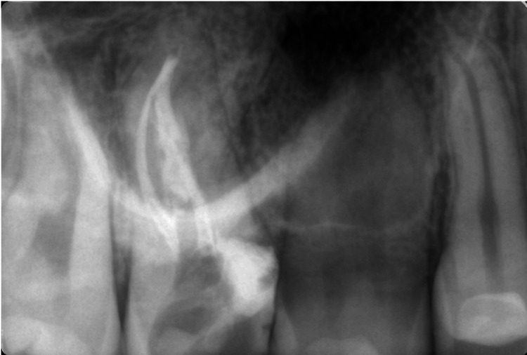
Root canal system variations can occur in each tooth group and significantly influence the outcome of the endodontic treatment. Upper first molars often present with some variations, mostly due to the presence of a second mesio-buccal root canal. Other types of atypical root canal system anatomy in upper first molars have also been reported but with a significantly smaller frequency. The aim of this article is to describe a clinical case of a C-shaped root canal configuration in a maxillary first molar - the diagnosis, preparation, irrigation, and final definitive obturation of the root canal system.
Keywords: c-shape; endodontic treatment; maxillary first molar; root canal system configuration; root canal system variation.
Copyright © 2025, Georgieva et al.
Conflict of interest statement
Human subjects: Consent for treatment and open access publication was obtained or waived by all participants in this study. Conflicts of interest: In compliance with the ICMJE uniform disclosure form, all authors declare the following: Payment/services info: All authors have declared that no financial support was received from any organization for the submitted work. Financial relationships: All authors have declared that they have no financial relationships at present or within the previous three years with any organizations that might have an interest in the submitted work. Other relationships: All authors have declared that there are no other relationships or activities that could appear to have influenced the submitted work.
Figures


Similar articles
-
[Root canal therapy of maxillary molars with atypical canals: A report of three cases].Beijing Da Xue Xue Bao Yi Xue Ban. 2024 Feb 18;56(1):190-195. doi: 10.19723/j.issn.1671-167X.2024.01.030. Beijing Da Xue Xue Bao Yi Xue Ban. 2024. PMID: 38318917 Free PMC article. Chinese.
-
Root canal treatment in an unusual maxillary first molar: a case report.Int Endod J. 2001 Dec;34(8):649-53. doi: 10.1046/j.1365-2591.2001.00445.x. Int Endod J. 2001. PMID: 11762503
-
CBVT analysis of canal configuration of the mesio-buccal root of maxillary first permanent molar teeth: An in vitro study.Contemp Clin Dent. 2012 Jul;3(3):277-81. doi: 10.4103/0976-237X.103618. Contemp Clin Dent. 2012. PMID: 23293481 Free PMC article.
-
C-shaped root canal in a maxillary first molar: a case report.Int Endod J. 2006 Feb;39(2):162-6. doi: 10.1111/j.1365-2591.2006.01069.x. Int Endod J. 2006. PMID: 16454798 Review.
-
Maxillary molars with morphologic variations of the palatal root canals: a report of four cases.J Endod. 2009 Jul;35(7):1060-5. doi: 10.1016/j.joen.2009.04.029. J Endod. 2009. PMID: 19567335 Review.
References
-
- Versiani MA, Basrani B, Sousa-Neto MD. Switzerland: Springer. Springer International Publishing; 2019. The Root Canal Anatomy in Permanent Dentition.
-
- Root canal anatomy of the human permanent teeth. Vertucci FJ. Oral Surg Oral Med Oral Pathol. 1984;58:589–599. - PubMed
-
- Root morphology of the maxillary first and second molars in an Iranian population using cone beam computed tomography. Ghoncheh Z, Zade BM, Kharazifard MJ. https://pubmed.ncbi.nlm.nih.gov/29167682/ J Dent (Tehran) 2017;14:115–122. - PMC - PubMed
-
- Root and root canal morphology of the human permanent maxillary first molar: a literature review. Cleghorn BM, Christie WH, Dong CC. J Endod. 2006;32:813–821. - PubMed
-
- Incidence and configuration of canal systems in the mesiobuccal root of maxillary first and second molars. Kulild JC, Peters DD. J Endod. 1990;16:311–317. - PubMed
Publication types
LinkOut - more resources
Full Text Sources