

Angioleiomyoma of cheek - A case report highlighting immunohistochemical diagnostic approach
- PMID: 40248625
- PMCID: PMC12002582
- DOI: 10.4103/jomfp.jomfp_138_24
Angioleiomyoma of cheek - A case report highlighting immunohistochemical diagnostic approach
Abstract
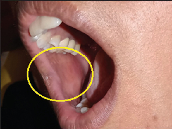
Benign smooth muscle tumours, known as leiomyomas, are comparatively frequent, with uterine cases accounting for 95% incidence. Oral leiomyomas typically appear as asymptomatic, slowly-growing submucosal lumps in the tongue, hard palate, or buccal mucosa. Three histologic kinds of leiomyomas are recognized: epithelioid leiomyoma, solid leiomyoma, and angioleiomyoma. The most prevalent type of leiomyomas affecting the oral cavity are solitary forms called angioleiomyomas, which typically develop in the subcutis. The diagnosis is commonly determined by histopathological and Immunohistochemistry (IHC) procedures. This case reports a 30-year-old female patient with a lesion on her right cheek region. The postsurgical specimen was routinely processed and stained with hematoxylin and eosin staining, and IHC studies confirmed the diagnosis of 'Benign spindle cell neoplasm-Angioleiomyoma'.
Keywords: Angioleiomyoma; benign spindle cell neoplasm; immunohistochemistry; leiomyoma.
Copyright: © 2025 Journal of Oral and Maxillofacial Pathology.
Conflict of interest statement
There are no conflicts of interest.
Figures


References
-
- Bhattacharyya I, Summerlin DJ, Cohen DM, Ellis GL, Bavitz JB, Gillham LL. Granular cell leiomyoma of the oral cavity. Oral Surg Oral Med Oral Pathol Oral Radiol Endod. 2006;102:353–9. - PubMed
-
- Hamid R, Chalkoo A, Tariq S, Bilal S, Wani S. Central angioleiomyoma of the mandible: A rare entity. J Cancer Res Ther. 2020;16:647–52. - PubMed
-
- Menditti D, Laino L, Nastri L, Caruso U, Fiore P, Baldi A. Oral angioleiomyoma: A rare pathological entity. In Vivo. 2012;26:161–3. - PubMed
-
- Heim S, Mandahl N, Kristoffersson U, Mitelman F, Rööser B, Rydholm A, et al. Structural chromosome aberrations in a case of angioleiomyoma. Cancer Genet Cytogenet. 1986;20:325–30. - PubMed
-
- Brooks JK, Nikitakis NG, Goodman NJ, Levy BA. Clinicopathologic characterization of oral angioleiomyomas. Oral Surg Oral Med Oral Pathol Oral Radiol Endod. 2002;94:221–7. - PubMed
Publication types
LinkOut - more resources
Full Text Sources