

Case Reports
doi: 10.5334/jbsr.3898.
eCollection 2025.
Endometriosis of the Round Ligaments in Twins: A Rare and Unique Presentation
Affiliations
- PMID: 40248836
- PMCID: PMC12005135
- DOI: 10.5334/jbsr.3898
Item in Clipboard
Case Reports
Endometriosis of the Round Ligaments in Twins: A Rare and Unique Presentation
J Belg Soc Radiol.
.
Abstract
Teaching point: The role of magnetic resonance imaging (MRI) in diagnosing endometriosis is growing, requiring radiologists to become familiar with both typical and atypical presentations of deep infiltrating endometriosis.
Keywords: Nuck; endometriosis; inguinal canal; magnetic resonance imaging; mri; twin; twins.
Copyright: © 2025 The Author(s).
Conflict of interest statement
The authors have no competing interests to declare.
Figures

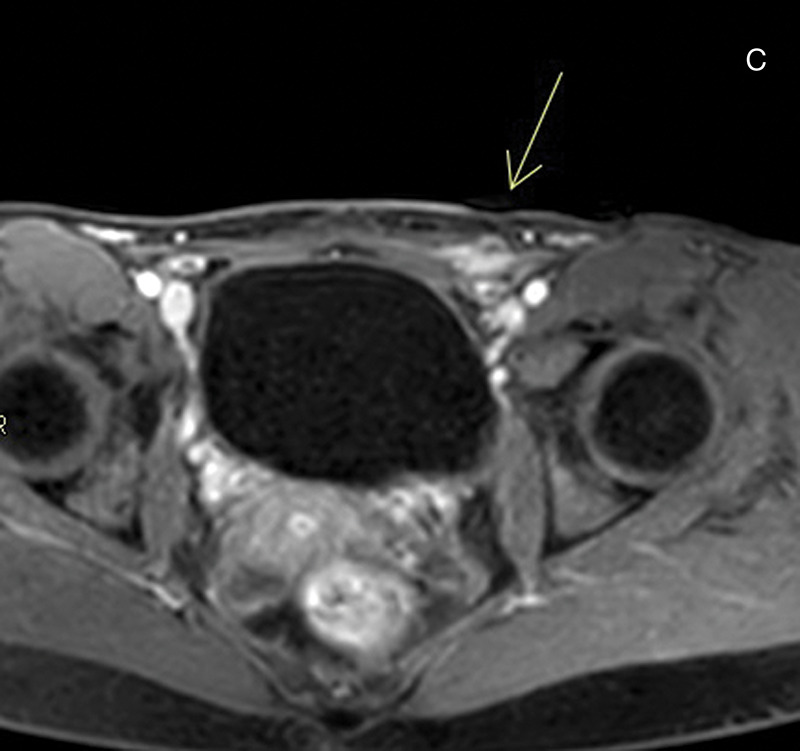
A. Ultrasound shows an mildly heterogeneous, hypoechoic mass with vascularity. B. Axial T1‑weighted fat suppressed image shows an isointense area in the left inguinal region. C. Same region as in Figure 1b, after intravenous administration of gadolinium, showing vivid lesional enhancement.

A. Ultrasound shows an mildly heterogeneous, hypoechoic mass with vascularity. B. Axial T1‑weighted fat suppressed image shows an isointense area in the left inguinal region. C. Same region as in Figure 1b, after intravenous administration of gadolinium, showing vivid lesional enhancement.
A. Ultrasound shows an mildly heterogeneous, hypoechoic mass with vascularity. B. Axial T1‑weighted fat suppressed image shows an isointense area in the left inguinal region. C. Same region as in Figure 1b, after intravenous administration of gadolinium, showing vivid lesional enhancement.
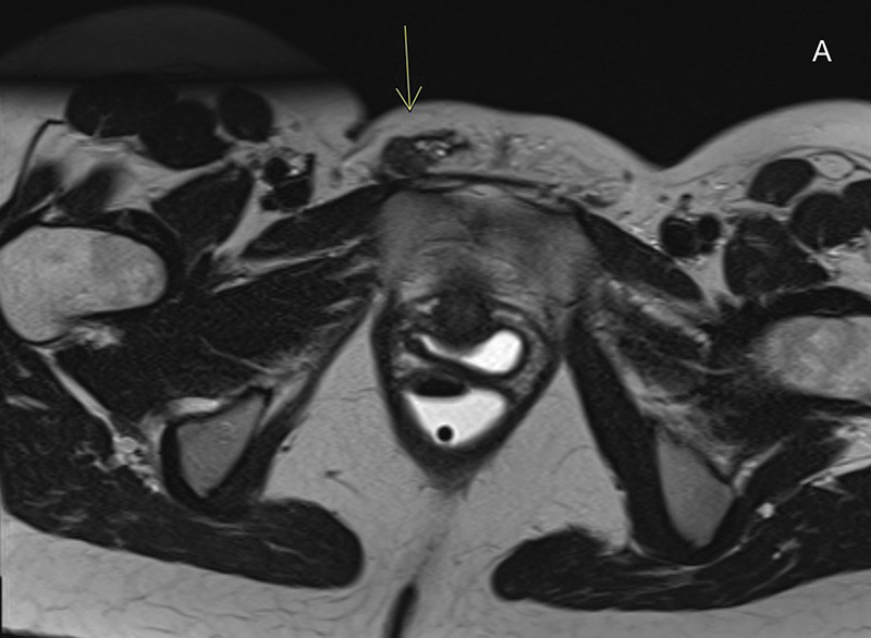
A. Axial T2‑weighted image shows a hypointense area in the right inguinal region with hyperintense foci. The mass is adjacent to the extrapelvic portion of the right round uterine ligament in the inguinal canal. B. Sagittal T2‑weighted image of the same area as Figure 2b. C. Axial T1‑weighted image with fat suppression shows the lesion with an isointense signal and discrete T1 hyperintense foci.

A. Axial T2‑weighted image shows a hypointense area in the right inguinal region with hyperintense foci. The mass is adjacent to the extrapelvic portion of the right round uterine ligament in the inguinal canal. B. Sagittal T2‑weighted image of the same area as Figure 2b. C. Axial T1‑weighted image with fat suppression shows the lesion with an isointense signal and discrete T1 hyperintense foci.

A. Axial T2‑weighted image shows a hypointense area in the right inguinal region with hyperintense foci. The mass is adjacent to the extrapelvic portion of the right round uterine ligament in the inguinal canal. B. Sagittal T2‑weighted image of the same area as Figure 2b. C. Axial T1‑weighted image with fat suppression shows the lesion with an isointense signal and discrete T1 hyperintense foci.
Similar articles
-
Hidden Depths: Unveiling Endometriosis in the Canal of Nuck.Cureus. 2024 Jul 20;16(7):e64975. doi: 10.7759/cureus.64975. eCollection 2024 Jul. Cureus. 2024. PMID: 39161483 Free PMC article.
-
Endometriosis in the canal of Nuck: Atypical manifestations in an unusual location.Can J Plast Surg. 2004 Summer;12(2):73-5. doi: 10.1177/229255030401200202. Can J Plast Surg. 2004. PMID: 24115879 Free PMC article.
-
Vulvar endometriosis and Nuck canal.Ann Ital Chir. 2014 Dec 29;85(ePub):S2239253X14023482. Ann Ital Chir. 2014. PMID: 25707680
-
Deep pelvic endometriosis: don't forget round ligaments. Review of anatomy, clinical characteristics, and MR imaging features.Abdom Imaging. 2014 Jun;39(3):622-32. doi: 10.1007/s00261-014-0091-3. Abdom Imaging. 2014. PMID: 24557639 Review.
-
Anterior pelvic endometriosis: MRI features.Abdom Imaging. 2010 Dec;35(6):742-9. doi: 10.1007/s00261-010-9600-1. Abdom Imaging. 2010. PMID: 20169439 Review.
Cited by
-
Nuck canal cyst: use of immunohistochemistry to reveal vestigial endometriosis hidden within mural granulation tissue.J Surg Case Rep. 2025 Aug 20;2025(8):rjaf540. doi: 10.1093/jscr/rjaf540. eCollection 2025 Aug. J Surg Case Rep. 2025. PMID: 40842487 Free PMC article.
References
-
- Adedipe T, Chukwujama U. Vulvar endometriosis mimicking as primary vulvodynia in a young nulliparous woman: Algorithm of care following a rapid literature review. J Clin Gynecol Obstet. 2021;10(2):35–39. 10.14740/jcgo735. - DOI
Publication types
LinkOut - more resources
Full Text Sources
