. 2025 Jul;56(7):1872-1882.
doi: 10.1161/STROKEAHA.124.048964. Epub 2025 Apr 18.

Hypoxic Preconditioning Enhances the Potential of Mesenchymal Stem Cells to Treat Neonatal Hypoxic-Ischemic Brain Injury

Affiliations

Hypoxic Preconditioning Enhances the Potential of Mesenchymal Stem Cells to Treat Neonatal Hypoxic-Ischemic Brain Injury

Sara T De Palma et al. Stroke. 2025 Jul.

Abstract

Background: Neonatal hypoxic-ischemic (HI) brain injury is one of the leading causes of long-term neurological morbidity in newborns. Current treatment options for HI brain injury are limited, but mesenchymal stem cell (MSC) therapy is a promising strategy to boost neuroregeneration after injury. Optimization strategies to further enhance the potential of MSCs are under development. The current study aimed to test the potency of hypoxic preconditioning of MSCs to enhance the therapeutic efficacy in a mouse model of neonatal HI injury.

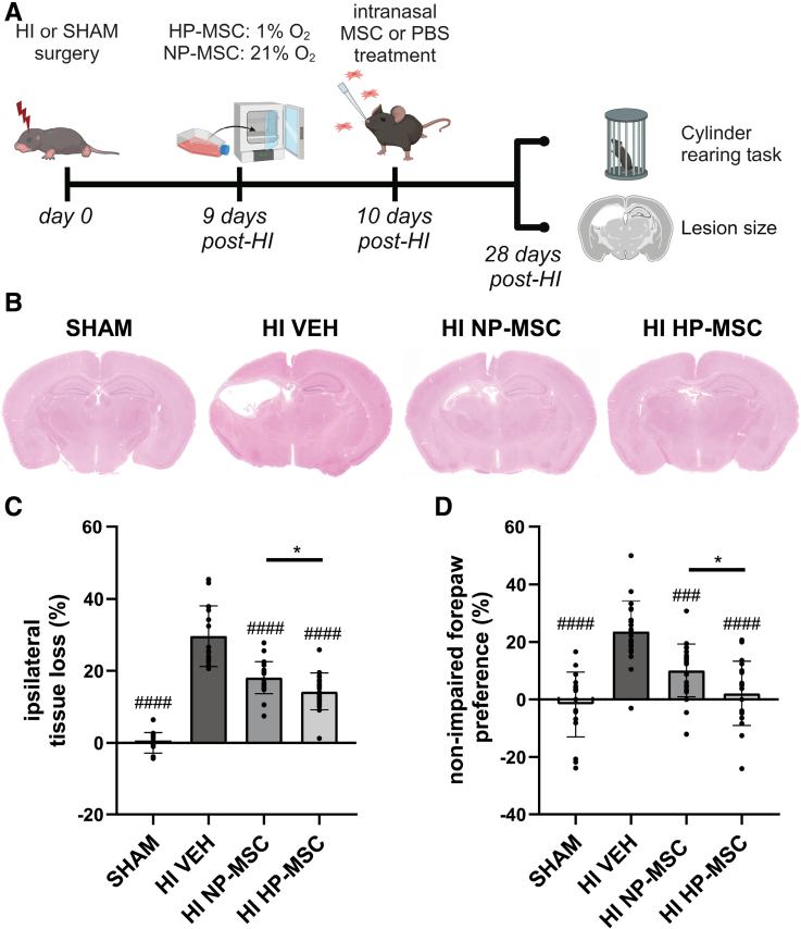
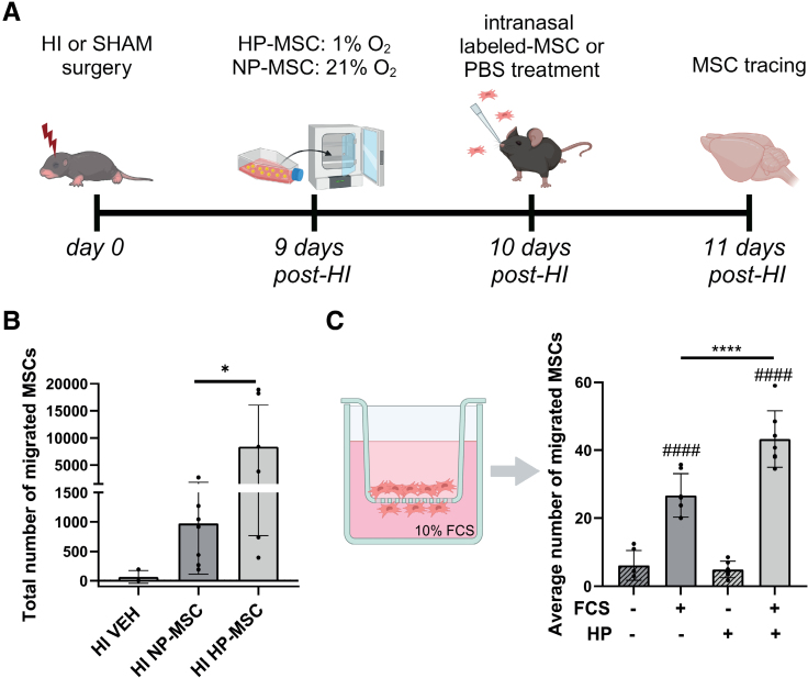
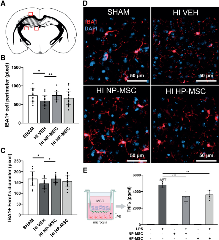
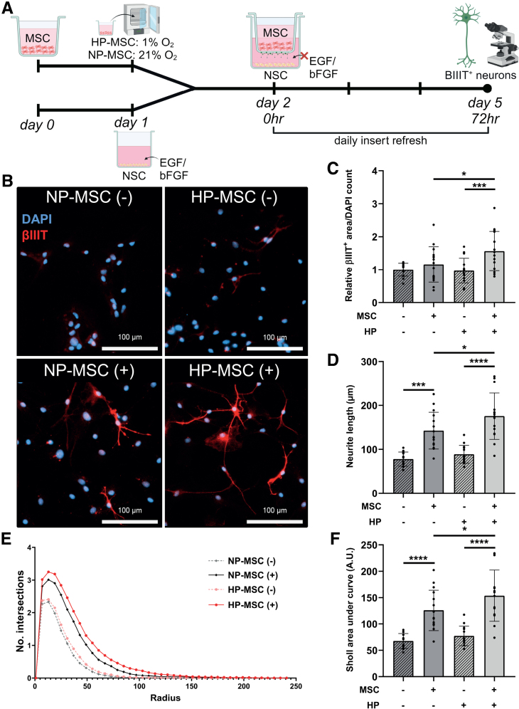
Methods: HI was induced on postnatal day 9 in C57Bl/6 mouse pups. MSCs were cultured under hypoxic (hypoxic-preconditioned MSCs [HP-MSCs], 1% O2) or normoxic-control (normoxic-preconditioned MSCs [NP-MSCs], 21% O2) conditions for 24 hours before use. At 10 days after HI, HP-MSCs, NP-MSCs, or vehicle were intranasally administered. Gold nanoparticle-labeled MSCs were used to assess MSC migration 24 hours after intranasal administration. At 28 days post-HI, lesion size, sensorimotor outcome, and neuroinflammation were assessed by hematoxylin and eosin staining, cylinder rearing task, and ionized calcium-binding adapter molecule 1 (IBA1) staining, respectively. In vitro, the effect of HP-MSCs was studied on transwell migration, neural stem cell differentiation and microglia activation, and the MSC intracellular proteomic content was profiled using quantitative Liquid Chromatography-Tandem Mass Spectrometry (LC-MS/MS).

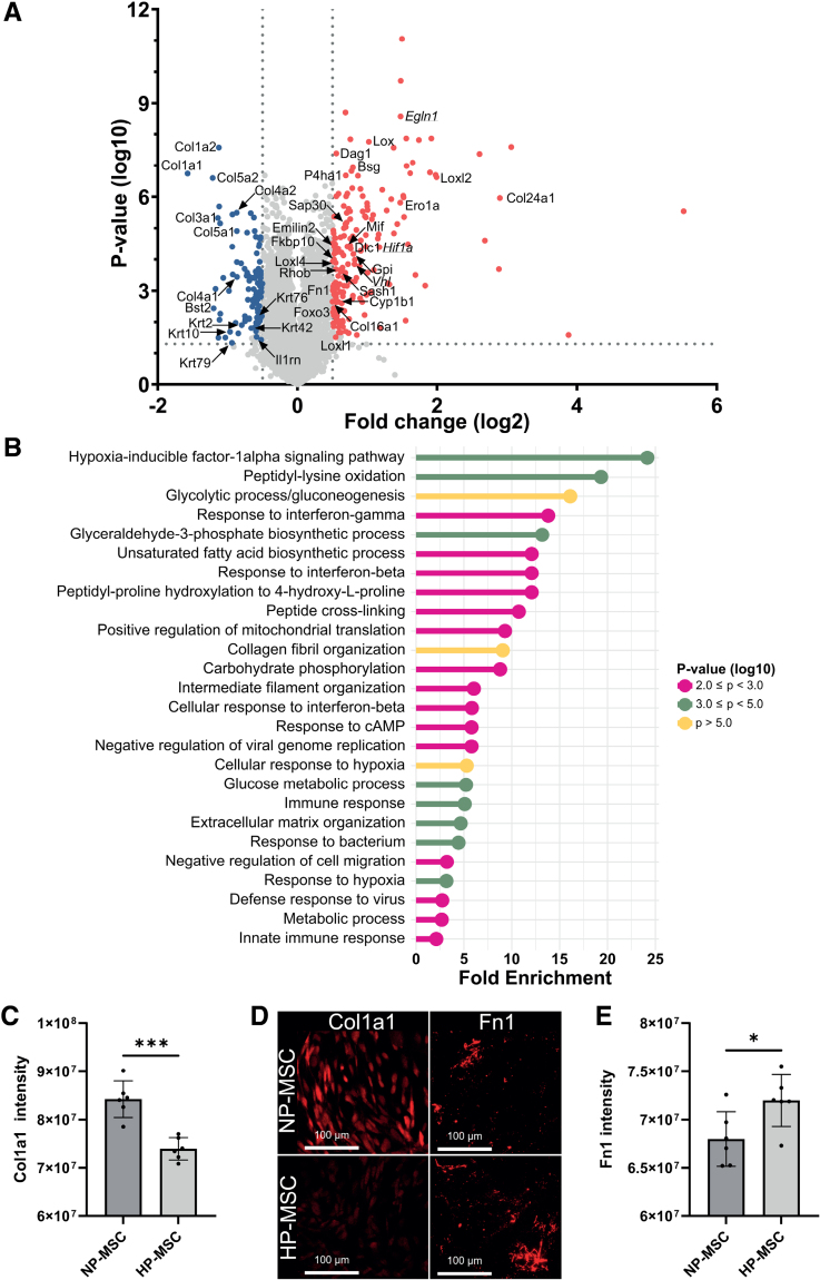
Results: Intranasally administered HP-MSCs were superior to NP-MSCs in reducing lesion size and sensorimotor impairments post-HI. Moreover, hypoxic preconditioning enhanced MSC migration in an in vitro set-up, and in vivo to the lesioned hemisphere after intranasal application. In addition, HP-MSCs enhanced neural stem cell differentiation into more complex neurons in vitro but had similar anti-inflammatory effects compared with NP-MSCs. Lastly, hypoxic preconditioning led to elevated abundances of proteins in MSCs related to extracellular matrix remodeling.

Conclusions: This study shows for the first time that hypoxic preconditioning enhanced the therapeutic efficacy of MSC therapy in a mouse model of neonatal HI brain injury by increasing the migratory and neuroregenerative capacity of MSCs.

Keywords: brain injuries; hypoxia-ischemia, brain; mesenchymal stem cells; neonate; neurogenesis; proteomics.


Conflict of interest statement

None.

Figures

Figure 1.
Figure 1.
Superior effect of hypoxic-preconditioned mesenchymal stem cells (HP-MSCs) on lesion size and functional outcome in a mouse model of neonatal hypoxic-ischemic (HI) brain injury. A, Overview of study design. B, Representative images of ipsilateral tissue loss visualized by hematoxylin and eosin staining in SHAM-control mice or HI-injured mice intranasally treated with either vehicle (VEH), normoxic-preconditioned MSCs (NP-MSCs), or HP-MSCs at 10 days post-HI. C, Quantification of ipsilateral tissue loss (%) at 28 days post-HI. D, Non-impaired forepaw preference at 28 days post-HI; SHAM: n=18, HI-VEH: n=21, HI–NP-MSC (C): n=23; NP-MSC (D): n=22, HI–HP-MSC: n=19. Data represent mean±SD. ###P<0.001, ####P<0.0001 significance relative to HI-VEH; *P<0.05 HI–HP-MSC vs HI–NP-MSC.
Figure 2.
Figure 2.
Enhanced migratory capacity of hypoxic-preconditioned mesenchymal stem cells (HP-MSCs) compared with normoxic-preconditioned MSCs (NP-MSCs) in vivo and in vitro. A, Overview of the study design. B, Gold-labeled MSC migration toward the ipsilateral lesioned hemisphere expressed in total number of cells; hypoxic-ischemic (HI)–vehicle (VEH): n=3, HI–NP-MSC: n=7, HI–HP-MSC: n=7. The HI-VEH group was used as negative control only and was not included in the statistical analysis. C, left, schematic overview of transwell migration assay; right, quantification of number of HP-MSCs or NP-MSCs migrated toward 10% fetal calf serum (FCS) in vitro; n=6–7 per condition. Data represent mean±SD. *P<0.05; ****P<0.0001 HP-MSCs vs NP-MSCs, ####P<0.0001 relative to no FCS condition.
Figure 3.
Figure 3.
Effect of hypoxic-preconditioned mesenchymal stem cells (HP-MSCs) on microglia activation after hypoxic-ischemic (HI) injury in vivo and lipopolysaccharide (LPS) exposure in vitro. A, Perilesional locations of images taken in ionized calcium-binding adapter molecule 1 (IBA1)-stained brain sections at P28. B, Quantification of perimeter of IBA1+ cells. C, Quantification of Feret diameter of IBA1+ cells. D, Representative fluorescent images of perilesional IBA1+ cells (40×) in SHAM controls, HI-vehicle (VEH), HI–normoxic-preconditioned MSC (NP-MSC), and HI–HP-MSC mice. E, left, schematic overview of non-contact co-culture of primary-isolated mouse microglia exposed to 50 ng/mL LPS with NP-MSCs or HP-MSC (purple cells) in the hanging insert, green dots represent MSC secretome that can reach the medium of the microglia; right, TNF-α (tumor necrosis factor-α) secretion by microglia after 24-hour exposure to LPS. Data represent mean±SD. B and C, SHAM: n=17, HI-VEH: n=19, HI–NP-MSC: n=22, and HI–HP-MSC: n=21. E, n=5 per condition. DAPI indicates 4',6-diamidino-2-phenylindole. *P<0.05, **P<0.01, ***P<0.001 between indicated groups, ####P<0.0001 compared with non-LPS condition.
Figure 4.
Figure 4.
Hypoxic-preconditioned mesenchymal stem cells (HP-MSCs) enhance differentiation of neural stem cells (NSCs) into more complex neurons than normoxic-preconditioned MSCs (NP-MSCs). A, Overview of experimental design. B, Representative fluorescent images (10×) of βIIIT (βIII-tubulin)+ cells (differentiated NSCs) in co-culture with NP-MSCs or HP-MSCs (+) or empty inserts (−). C, Quantification of βIIIT+ area normalized to the cell numbers (4',6-diamidino-2-phenylindole [DAPI] count) relative to the NP-MSCs (−) condition. D, Quantification of neurite length of βIIIT+ neurons. E, Number of intersections of βIIIT+ neurons with circles of increasing radius in Sholl analysis. F, Quantification of Sholl analysis by area under the curve. Data represent mean±SD. All: n=13–15 per condition out of 3 independent experiments. A.U. indicates arbitrary unit; bFGF, basic fibroblast growth factor; and EGF, epidermal growth factor. *P<0.05, ***P<0.001, and ****P<0.0001.
Figure 5.
Figure 5.
Hypoxic preconditioning of mesenchymal stem cells (MSCs) leads to increased intracellular expression of Hif1α (hypoxia-inducible factor 1 alpha) and proteins primarily involved in glucose metabolism and ECM (extracellular matrix) remodeling. A, Volcano plot with significantly upregulated (red) and downregulated (blue) proteins directly after hypoxic preconditioning. Annotated proteins are found to be involved in migratory processes by enrichment analysis. Hif1α and its interactors are underlined; n=2 per condition. B, Pathway enrichment analysis of upregulated and downregulated proteins in hypoxic-preconditioned MSCs (HP-MSCs) vs normoxic-preconditioned MSCs (NP-MSCs). C, Quantification of Col1a1 (collagen alpha-1(I) chain) expression by NP-MSCs and HP-MSCs. D, Representative fluorescent images (10×) of Fn1 (fibronectin 1)+ ECM and Col1a1+ MSCs in vitro. E, Quantification of Fn1 expression in the ECM of NP-MSCs and HP-MSCs. Data represent mean±SD. C and E, n=6 per condition. Bsg indicates basigin; Bst2, Bone marrow stromal antigen 2; Col16a1, collagen alpha-1(XVI) chain; Col1a1, collagen alpha-1(I) chain; Col1a2, collagen alpha-2(I) chain; Col24a1, collagen alpha-1(XXIV) chain; Col3a1, collagen alpha-1(III) chain; Col4a1, collagen alpha-1(IV) chain; Col4a2, collagen alpha-2(IV) chain; Col5a1, collagen alpha-1(V) chain; Col5a2, collagen alpha-2(V) chain; Cyp1b1, cytochrome P450 1B1; Dag1, dystroglycan 1; Dlc1, Rho GTPase-activating protein 7; Egln1, Egl [egg-laying defective] nine homolog 1; Ero1a, ERO1-like protein alpha; Fkbp10, peptidyl-prolyl cis-trans isomerase FKBP10; Foxo3, Forkhead box protein O3; Gpi, glucose-6-phosphate isomerase; Il1rn, interleukin-1 receptor antagonist protein; Krt10, keratin, type I cytoskeletal 10; Krt2, keratin, type II cytoskeletal 2 epidermal; Krt42, keratin, type I cytoskeletal 42; Krt5, keratin, type II cytoskeletal 5; Krt76, keratin, type II cytoskeletal 2 oral; Krt79, keratin, type II cytoskeletal 79; Lox, protein-lysine 6-oxidase; Loxl1, lysyl oxidase homolog 1; Loxl2, Lysyl oxidase homolog 2; Loxl4, Lysyl oxidase homolog 4; Mif, Macrophage migration inhibitory factor; P4ha1, prolyl 4-hydroxylase subunit alpha-1; Rhob, Rho-related GTP-binding protein RhoB; Sap30, histone deacetylase complex subunit SAP30; Sash1, SAM and SH3 domain-containing protein 1; and Vhl, Von Hippel–Lindau tumor suppressor. *P<0.05, ***P<0.001.

Similar articles

Cited by

References

    1. Lee AC, Kozuki N, Blencowe H, Vos T, Bahalim A, Darmstadt GL, Niermeyer S, Ellis M, Robertson NJ, Cousens S, et al. Intrapartum-related neonatal encephalopathy incidence and impairment at regional and global levels for 2010 with trends from 1990. Pediatr Res. 2013;74:50–72. doi: 10.1038/pr.2013.206 - PMC - PubMed
    1. Perez A, Ritter S, Brotschi B, Werner H, Caflisch J, Martin E, Latal B. Long-term neurodevelopmental outcome with hypoxic-ischemic encephalopathy. J Pediatr. 2013;163:454–459. doi: 10.1016/j.jpeds.2013.02.003 - PubMed
    1. Kariholu U, Montaldo P, Markati T, Lally P, Pryce R, Teiserskas J, Liow N, Oliveira V, Soe A, Shankaran S, et al. Therapeutic hypothermia for mild neonatal encephalopathy: a systematic review and meta-analysis. Arch Dis Child Fetal Neonatal Ed. 2020;105:F225–F228. doi: 10.1136/archdischild-2018-315711 - PubMed
    1. Baak LM, Wagenaar N, van der Aa NE, Groenendaal F, Dudink J, Tataranno ML, Mahamuud U, Verhage CH, Eijsermans RMJC, Smit LS, et al. Feasibility and safety of intranasally administered mesenchymal stromal cells after perinatal arterial ischaemic stroke in the Netherlands (PASSIoN): a first-in-human, open-label intervention study. Lancet Neurol. 2022;21:528–536. doi: 10.1016/S1474-4422(22)00117-X - PubMed
    1. Donega V, Van Velthoven CTJ, Nijboer CH, Van Bel F, Kas MJH, Kavelaars A, Heijnen CJ. Intranasal mesenchymal stem cells treatment for neonatal brain damage: long-term cognitive and sensorimotor improvement. PLoS One. 2013;8:e51253. doi: 10.1371/journal.pone.0051253 - PMC - PubMed