. 2025 Apr;286(4):e70047.
doi: 10.1002/jmor.70047.

Ontogeny of a Brazilian Late Triassic Traversodontid (Cynodontia, Cynognathia): Anatomical and Paleoecological Implications

Affiliations

Ontogeny of a Brazilian Late Triassic Traversodontid (Cynodontia, Cynognathia): Anatomical and Paleoecological Implications

Lívia Roese-Miron et al. J Morphol. 2025 Apr.

Abstract

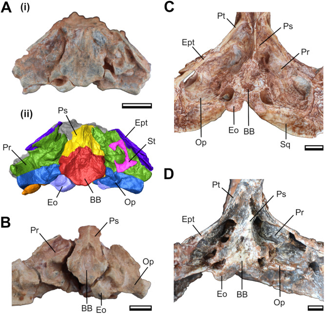
Investigating the developmental patterns of extinct species provides valuable insights into their anatomy, biology and ecomorphological adaptations. Research on the ontogeny of non-mammaliaform cynodonts has offered significant contributions to our understanding of these aspects. Here, we aim to describe and discuss the intraspecific and ontogenetic variation of the skull of the Brazilian traversodontid Siriusgnathus niemeyerorum (Candelária Sequence, Upper Triassic). We evaluated an ontogenetic series of the species through qualitative comparison and allometric analyses using cranial measures. Our findings reveal several trends during skull growth, including a relative increase in rostrum length, a relative decrease in orbit size, and changes in the zygomatic arch and temporal fenestra proportions. These patterns, when analyzed in the context of the adductor musculature, may be correlated with changes in feeding behaviour, similar to those described for the gomphodontosuchine Exaeretodon argentinus. We also report changes in cranial ornamentation, bone fusion, and suture complexity throughout ontogeny. Overall, this study provides a greater understanding of the cranial ontogenetic patterns of S. niemeyerorum, contributing to the knowledge of its intraspecific variation. The possible ecological implications of these findings highlight the importance of ontogenetic studies for elucidating the biology of extinct taxa.

Keywords: Candelária sequence; Siriusgnathus niemeyerorum; intraspecific variation; ontogeny; skull anatomy.


Conflict of interest statement

The authors declare no conflicts of interest.

Figures

Figure 1
Figure 1
Measurements of the Siriusgnathus niemeyerorum specimens. Reconstruction of the cranium in (A) dorsal view; (B) ventral view; and (C) lateral view. BL, Basicranium Length; BSL, Basal Skull Length; BW, Basicranium Width; IW, Interorbital Width; NTL, Non‐temporal Length; OL, Orbit Length; OW, Orbit Width; PL, Palate Length; RL, Rostrum Length; RW, Rostrum Width; SSW, Suborbital Skull Width; SW, Skull Width; TFL, Temporal Fenestra Length; TFW, Temporal Fenestra Width; TL1, Temporal Length 1 (until the posterior limit of the sagittal crest); TL2, Temporal Length 2 (until the posterior limit of the zygomatic arch); TPW, Transverse Process Width; TSL, Total Skull Length; UDL, Upper Diastema Length (between canine and first postcanine); UPPD, Upper Posterior Postcanine Distance; UPRL, Upper Postcanine Row Length; ZAH, Zygomatic Arch Height; ZAL, Zygomatic Arch Length.
Figure 2
Figure 2
Linear regressions of cranium measurements of Siriusgnathus niemeyerorum exhibiting the relationships between: (A) total skull lenght and basal skull length; (B) basal skull length and skull width; (C) basal skull length and suborbital skull width; (D) skull width and suborbital skull width; (E) suborbital skull width and basicranium length; (F) suborbital skull width and basicranium width; (G) basal skull length and rostrum length; and (H) basal skull length and temporal length. The number of specimens included varies (5–8) based on which measurements were possible. All axes are log‐transformed. The statistics (slope, intercept, R 2 and p value) are included in each plot.
Figure 3
Figure 3
Linear regressions of cranium measurements of Siriusgnathus niemeyerorum exhibiting the relationships between: (A) basal skull length and orbit width; (B) basal skull length and zygomatic arch height; (C) basal skull length and temporal fenestra length; (D) basal skull length and temporal fenestra width; (E) basal skull length and palate length; (F) basal skull length and transverse process width; (G) basal skull length and upper posterior postcanine distance; and (H) basal skull length and upper postcanine row length. The number of specimens included varies (5–8) based on which measurements were possible. All axes are log‐transformed. The statistics (slope, intercept, R 2 and p value) are included in each plot.
Figure 4
Figure 4
Crania of Siriusgnathus niemeyerorum in dorsal view, in ascending order of size. (A) CAPPA/UFSM 0191; (B) CAPPA/UFSM 0124; (C) CAPPA/UFSM 0329; (D) CAPPA/UFSM 0394; (E) CAPPA/UFSM 0125; (F) CAPPA/UFSM 0032; (G) CAPPA/UFSM 0330; (H) CAPPA/UFSM 0109. Scale bars = 30 mm.
Figure 5
Figure 5
Crania of Siriusgnathus niemeyerorum in ventral view, in ascending order of size. (A) CAPPA/UFSM 0191; (B) CAPPA/UFSM 0124; (C) CAPPA/UFSM 0329; (D) CAPPA/UFSM 0394; (E) CAPPA/UFSM 0125; (F) CAPPA/UFSM 0032; (G) CAPPA/UFSM 0330; (H) CAPPA/UFSM 0109. Scale bars = 30 mm.
Figure 6
Figure 6
Crania of Siriusgnathus niemeyerorum in lateral view, in ascending order of size. (A) CAPPA/UFSM 0191 (lateral right, mirrored); (B) CAPPA/UFSM 0124 (lateral left); (C) CAPPA/UFSM 0329 (lateral left); (D) CAPPA/UFSM 0394 (lateral right, mirrored); (E) CAPPA/UFSM 0125 (lateral left); (F) CAPPA/UFSM 0032 (lateral right, mirrored); (G) CAPPA/UFSM 0330 (lateral right, mirrored); (H) CAPPA/UFSM 0260 (lateral right, mirrored); (I) CAPPA/UFSM 0109 (lateral left). Scale bars = 30 mm.
Figure 7
Figure 7
Crania of Siriusgnathus niemeyerorum in occipital view, in ascending order of size. (A) CAPPA/UFSM 0074; (B) CAPPA/UFSM 0191; (C) CAPPA/UFSM 0329; (D) CAPPA/UFSM 0394; (E) CAPPA/UFSM 0125; (F) CAPPA/UFSM 0032; (G) CAPPA/UFSM 0330; (H) CAPPA/UFSM 0109. Scale bars = 20 mm.
Figure 8
Figure 8
Lower jaw of Siriusgnathus niemeyerorum. (A) CAPPA/UFSM 0334 in lateral right (i), and lateral left (ii) views; (B) CAPPA/UFSM 0261 in lateral right (i) and lateral left (ii) views; (C) CAPPA/UFSM 0125 in lateral left (i) and medial left (ii) views; (D) CAPPA/UFSM 0032 in lateral right (i) and medial right (ii) views. Scale bars = 20mm (A–C) and 30mm (D).
Figure 9
Figure 9
Basicranial anatomy of Siriusgnathus niemeyerorum in ventral view. (A) CAPPA/UFSM 0074: photograph (i) and 3D digital models (ii); (B) CAPPA/UFSM 0103; (C) CAPPA/UFSM 0329; (D) CAPPA/UFSM 0032. Scale bars = 10 mm. BB, basisphenoid + basioccipital; Eo, exoccipital; Ept, epipterygoid; Op, opisthotic; Pr, prootic; Ps, parasphenoid; Pt, pterygoid; Sq, squamosal; St, stapes.
Figure 10
Figure 10
Hypothetical reconstruction of the ontogenetic trajectory of the cranium of Siriusgnathus niemeyerorum. (A) juvenile, anatomy based on CAPPA/UFSM 0191 and 0124. (B) young adult, anatomy based CAPPA/UFSM 0329 (sutures based on Roese‐Miron et al. in review). (C) old adult, anatomy based on CAPPA/UFSM 0032, 0109 and 0330. (A–C) are not to scale. (D) outlines to scale of the three hypothetical specimens depicted in (A–C) based on the size of CAPPA/UFSM 0191, CAPPA/UFSM 0329 and CAPPA/UFSM 0330, respectively. i, elongation of the rostrum; ii, increase in rostrum width; iii, decrease in orbit relative size; iv, increase in interorbital width; v, lateral development of the posterior portion of the zygomatic arches; vi, increase in cranial ornamentation; vii, increase in suture complexity.
Figure 11
Figure 11
Artistic representation of three individuals of Siriusgnathus niemeyerorum in a Late Triassic landscape of southern Brazil. The adult (on the left) is followed by two juveniles. Artwork by Márcio L. Castro.

References

    1. Abdala, F. , Barberena M. C., and Dornelles J.. 2002. “A New Species of the Traversodontid Cynodont Exaeretodon From the Santa Maria Formation (Middle/Late Triassic) of Southern Brazil.” Journal of Vertebrate Paleontology 22, no. 2: 313–325.
    1. Abdala, F. , and Damiani R.. 2004. “Early Development of the Mammalian Superficial Masseter Muscle in Cynodonts.” Palaeontologica Africana 40: 23–29.
    1. Abdala, F. , and Gaetano L. C.. 2017. “The Late Triassic Record of Cynodonts: Time of Innovations in the Mammalian Lineage.” In Late Triassic World: Earth in a Time of Transition, edited by Tanner L. H., 407–445. Springer.
    1. Abdala, F. , Gaetano L. C., Martinelli A. G., Soares M. B., Hancox P. J., and Rubidge B. S.. 2020. “Non‐Mammaliaform Cynodonts From Western Gondwana and the Significance of Argentinean Forms in Enhancing Understanding of the Group.” Journal of South American Earth Sciences 104: 102884.
    1. Abdala, F. , and Giannini N. P.. 2000. “Gomphodont Cynodonts of the Chañares Formation: The Analysis of an Ontogenetic Sequence.” Journal of Vertebrate Paleontology 20, no. 3: 501–506.

LinkOut - more resources