. 2025 Jun 1;214(6):1236-1246.
doi: 10.1093/jimmun/vkaf042.

Innate immune sensor NLRP3 drives PANoptosome formation and PANoptosis

Affiliations

Innate immune sensor NLRP3 drives PANoptosome formation and PANoptosis

Bhesh Raj Sharma et al. J Immunol. .

Abstract

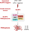
Inflammasomes are multiprotein innate immune complexes formed in response to infections, tissue damage, or cellular stress that promote the maturation and release of IL-1β/IL-18 and are implicated in lytic cell death. The NLRP3 inflammasome is canonically activated by an initial priming event followed by an activation stimulus, leading to rapid cell death that occurs through caspase-1 (CASP1) and gasdermin D (GSDMD) activation, called pyroptosis. CASP1- and GSDMD-deficient cells are protected from the rapid LPS plus ATP-induced pyroptosis. However, innate immune responses physiologically occur over time, extending beyond minutes to hours and days. Therefore, in this study, we assessed lytic cell death beyond the early timepoints. While cells lacking the innate immune sensor NLRP3 were protected from cell death induced by the canonical NLRP3 trigger, LPS priming and ATP stimulation (LPS plus ATP), for extended time, CASP1- and GSDMD-deficient cells started to lyse in a time-dependent manner after 2 h. Nevertheless, robust IL-1β and IL-18 release was still dependent on CASP1 activation. These data suggested that NLRP3 engages an additional innate immune, lytic cell death pathway. Indeed, LPS plus ATP induced the activation of caspases and RIPKs associated with PANoptosis in WT cells, and cells deficient in PANoptosis machinery were protected from cell death for extended times. A PANoptosome complex containing NLRP3, ASC, CASP8, and RIPK3 was observed by microscopy in WT, as well as CASP1- or GSDMD-deficient, cells by 30 min post-stimulation. Overall, these findings highlight the central role of NLRP3 as a PANoptosome sensor. Given the physiological role of innate immune cell death, PANoptosis, in health and disease, our study emphasizes the importance of a comprehensive understanding of PANoptosomes, and their components, as therapeutic targets.

Keywords: PANoptosome; RIPK; caspase; inflammasome; inflammation.


Conflict of interest statement

T.-D.K. was a consultant for Pfizer.

Figures
None
Graphical abstract
Figure 1.
Figure 1.
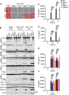
NLRP3 drives lytic cell death independent of CASP1 or GSDMD. (A) Representative images of cell death are shown for bone marrow-derived macrophages (BMDMs) at 0.5 h and 6 h post-treatment with LPS plus ATP. (B) Immunoblot analysis of pro- (P45) and activated (P20) caspase-1 (CASP1), pro- (P53) and activated (P30) gasdermin D (GSDMD), pro- (P53) and activated (P34) gasdermin E (GSDME), pro- (P55) and cleaved (P44 and P18) caspase-8 (CASP8), pro- (P35) and cleaved (P20) caspase-7 (CASP7), pro- (P35) and cleaved (P19 and P17) caspase-3 (CASP3), phosphorylated MLKL (pMLKL), total MLKL (tMLKL), HMGB1, and LDH in wild type (WT), Nlrp3–/–, Asc–/–, Casp1–/–, and Gsdmd–/– BMDMs at 0.5 h and 6 h post-treatment with LPS plus ATP. GAPDH was used as a loading control. Sup indicates supernatant. Blots for pMLKL and tMLKL were performed on the same membrane, with stripping and reprobing as described in the methods. (C–F) Pro-inflammatory cytokines released from BMDMs at 0.5 h and 6 h post-treatment with LPS plus ATP. Data are representative of at least 3 independent biological replicates (A–F). The scale bar is representative of 50 μm (A). ****P < 0.0001; ns, not statistically significant. Two-way ANOVA (C–F) was used. The data are represented as mean ± SEM in C–F.
Figure 2.
Figure 2.
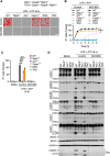
CASP8 acts redundantly with CASP1 to drive NLRP3-dependent PANoptosis. (A) Representative images of cell death are shown at 6 h post-treatment with LPS plus ATP with or without MCC950 treatment in wild type (WT), Nlrp3–/–, Asc–/–, Casp1–/–, Casp8–/–Ripk3–/– (referred to as DKO), and Casp1–/–Casp8–/–Ripk3–/– (referred to as TKO) bone marrow-derived macrophages (BMDMs). MCC950 was added 30 min before ATP. (B) Real time analysis of cell death in WT, Nlrp3–/–, Asc–/–, Casp1–/–, DKO, and TKO BMDMs in response to LPS plus ATP with or without MCC950 treatment. (C) Quantification of cell death in WT, Nlrp3–/–, Asc–/–, Casp1–/–, DKO, and TKO BMDMs at 6 h post-treatment with LPS plus ATP with or without MCC950 treatment. (D) Immunoblot analysis of pro- (P45) and activated (P20) caspase-1 (CASP1), pro- (P53) and activated (P30) gasdermin D (GSDMD), pro- (P53) and activated (P34) gasdermin E (GSDME), pro- (P55) and cleaved (P44 and P18) caspase-8 (CASP8), pro- (P35) and cleaved (P20) caspase-7 (CASP7), pro- (P35) and cleaved (P19 and P17) caspase-3 (CASP3), phosphorylated MLKL (pMLKL), total MLKL (tMLKL), HMGB1, and LDH in WT, Nlrp3–/–, Asc–/–, DKO, and TKO BMDMs at 6 h post-treatment with LPS plus ATP with or without MCC950 treatment. Blots for CASP3 and CASP7 were performed on the same membrane, with stripping and reprobing as described in the methods. GAPDH was used as a loading control. Sup indicates supernatant. Data are representative of at least three independent biological replicates (A–D). The scale bar is representative of 50 μm (A). *P < 0.05; ***P < 0.001; ****P < 0.0001. Two-way ANOVA (B, C) was used. The data are represented as mean ± SEM in B, C.
Figure 3.
Figure 3.
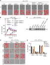
Blocking PANoptosis protects against LPS plus ATP-induced cell death. (A) Representative images of cell death in wild type (WT), Nlrp3–/–, Gsdmd–/–, Gsdme–/–, Mlkl–/–, Gsdmd–/–Gsdme–/–, Gsdmd–/–Gsdme–/–Mlkl–/–, and Ninj1–/– bone marrow-derived macrophages (BMDMs) at 0 h or 6 h post-treatment with LPS plus ATP. (B) Real-time analysis of cell death in WT, Nlrp3–/–, Gsdmd–/–, Gsdme–/–, Mlkl–/–, Gsdmd–/–Gsdme–/–, Gsdmd–/–Gsdme–/–Mlkl–/–, and Ninj1–/– BMDMs in response to LPS plus ATP. (C) Immunoblot analysis of LDH in the supernatant from Gsdme–/–, Mlkl–/–, Gsdmd–/–Gsdme–/–, Gsdmd–/–Gsdme–/–Mlkl–/–, and Ninj1–/– BMDMs at 6 h post-treatment with LPS plus ATP. (D, E) Representative images of cell death (D) and quantification of cell death (E) in WT, Nlrp3–/–, Casp1–/–, and Gsdmd–/– BMDMs at 6 h post-treatment with LPS plus ATP with or without the pan-caspase inhibitor zVAD and MLKL inhibitor (MLKLi). zVAD and MLKLi were added 1 h before ATP. The data are representative of at least 3 independent biological replicates (A–E). The scale bar is representative of 50 μm (A, D).
Figure 4.
Figure 4.
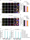
NLRP3-PANoptosome containing ASC, CASP8, and RIPK3 forms in response to canonical NLRP3 triggers. (A–F) Wild type (WT), Casp1–/–, and Gsdmd–/– bone marrow-derived macrophages (BMDMs) were unstimulated (0 h) or stimulated with LPS plus ATP (A, B, E) or LPS plus nigericin (C, D, F) for 0.5 h and stained for NLRP3, ASC, caspase-8 (CASP8), and RIPK3, and counter-stained with DAPI to visualize nuclei. (A, C) Representative images of cells containing co-localized NLRP3, ASC, CASP8, and RIPK3 are shown. The magnified view of the boxed area (merged) is shown on the right (enlarged). Scale bar = 5 µm (Merge with DAPI) and 1 µm (Enlarged). (B, D) Quantification showing the percentage of cells with ASC+NLRP3+CASP8+RIPK3+ specks out of the total population of cells with ASC+ specks (n ≥ 100) in WT, Casp1–/–, and Gsdmd–/– BMDMs at 0.5 h post-stimulation with LPS plus ATP (B) or LPS plus nigericin (D). (E, F) Compositional analysis of ASC specks (n ≥ 100) in WT, Casp1−/−, and Gsdmd−/− BMDMs at 0.5 h post-LPS plus ATP (E) or LPS plus nigericin (F) stimulation. Data are representative of at least 3 independent biological replicates (A–F). The data are represented as mean ± SEM (B, D–F).

References

    1. Sundaram B, Tweedell RE, Prasanth Kumar S, Kanneganti TD.The NLR family of innate immune and cell death sensors. Immunity. 2024;57:674–699. - PMC - PubMed
    1. Kanneganti TD et al. Bacterial RNA and small antiviral compounds activate caspase-1 through cryopyrin/Nalp3. Nature. 2006;440:233–236. - PubMed
    1. Man SM, Kanneganti TD. Converging roles of caspases in inflammasome activation, cell death and innate immunity. Nat Rev Immunol. 2016;16:7–21. - PMC - PubMed
    1. Martinon F, Burns K, Tschopp J. The inflammasome: a molecular platform triggering activation of inflammatory caspases and processing of proIL-beta. Mol Cell. 2002;10:417–426. - PubMed
    1. Franchi L et al. Cytosolic flagellin requires Ipaf for activation of caspase-1 and interleukin 1beta in salmonella-infected macrophages. Nat Immunol. 2006;7:576–582. - PubMed

MeSH terms

LinkOut - more resources