. 2025 Apr 18;11(16):eadu0975.
doi: 10.1126/sciadv.adu0975. Epub 2025 Apr 18.

Hippocampal perineuronal net degradation identifies prefrontal and striatal circuits involved in schizophrenia-like changes in marmosets

Affiliations

Hippocampal perineuronal net degradation identifies prefrontal and striatal circuits involved in schizophrenia-like changes in marmosets

Miriam A Gwilt et al. Sci Adv. .

Abstract

In schizophrenia, anterior hippocampus (aHipp) overactivity is associated with orbitofrontal cortex (OFC) dysfunction, but the contribution to symptomatology is unknown. In rodents, degradation of the hippocampal perineuronal net (PNN) replicates this overactivity, but uncertainty over rodent/human prefrontal homology limits translation to humans. Here, we test the hypothesis that aHipp PNN degradation in a species with a human-like prefrontal cortex, the marmoset, alters aHipp-striatal and aHipp-OFC circuitry. Microdialysis and [18F]-fluoro-l-dihydroxyphenylalanine positron emission tomography identified increased dopamine synthesis in the associative striatum, but not the nucleus accumbens, as is seen in schizophrenia, and elevated dopamine and noradrenaline in the OFC. Behaviorally, activity was elevated in a marmoset version of the amphetamine-induced activity test, and impaired probabilistic discrimination learning was seen in an OFC/striatum-dependent task that computational modeling suggests was due to loss of goal-directed behavior. Together, these findings demonstrate that a loss of primate aHipp PNNs is sufficient to induce striatal and prefrontal dysfunction consistent with that observed in humans with schizophrenia.


Figures
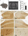
Fig. 1.
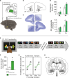
Fig. 1.. PNNs were selectively degraded throughout the aHipp.
(A) Schematic depicting bilateral cannulae targeting the aHipp. (B) Representative MRI of bilateral double cannulae targeting the aHipp. (C) Quantification of PNN staining after unilateral chondroitinase ABC digestion. The ratio of PNN staining in the degraded hippocampus (black) as a fraction of all staining is reduced when compared to the intact hippocampus (white). Means ± SEM; *P < 0.05. (D to I) PNNs (brown) surround PV-positive interneurons (gray) throughout the aHipp. Photomicrographs at increasing levels of magnification (from top to bottom) in an intact [(D), (F), and (H)] and degraded [(E), (G), and (I)] hippocampus demonstrate the loss of PNNs in the latter. In each case, the area enclosed in the rectangle is enlarged in the image immediately below. Panels (H) and (I) specifically demonstrate the loss of PNNs from around PV-positive cells.
Fig. 2.
Fig. 2.. aHipp PNN degradation increases activity but does not alter extracellular dopamine levels in the Acb.
(A) Bilateral aHipp PNN degradation amplified the stereotypical grooming behavior observed after intramuscular amphetamine (0.5 mg/kg; n = 4) treatment; ***P < 0.001. Means ± SEM. (B) Amphetamine did not affect locomotor activity, but aHipp PNN degradation alone was sufficient to increase the mean locomotor activity observed after saline injection across all time bins. Inset: mean locomotor activity before (white bar) and after (black bar) PNN degradation and saline treatment (n = 4); *P < 0.05. Means ± SEM. (C) Schematic depicting the experimental Acb microdialysis procedure. (D) Schematic depicting the location of the dialysis probes within the Acb of each animal, as determined by postmortem histology, plotted here on a single coronal section (AP 11 to 12.2). For histological schematics of the dialysis subjects, see fig. S2. (E) Representative histological section, with arrows marking the small holes at the position of the cannula tips. (F) Extracellular dopamine and noradrenaline levels within the Acb under tonic baseline (BL) conditions and phasic conditions (evoked by 75 mM K+; K+) were unchanged by aHipp PNN degradation (n = 4; means ± SEM).
Fig. 3.
Fig. 3.. aHipp PNN degradation increases dopamine synthesis in the associative striatum.
(B and D) Coronal co-registered MRI and −log10(P value) maps depicting the significance of regional increases in k3s, the rate at which [18F]-DOPA is decarboxylated to [18F]-fluorodopamine. Depicted at AP 7.5 (B) and AP 6.5 (D) alongside matched region of interest schematics (A and C), respectively (green, GPexternal; yellow, GPinternal; red, caudate tail). Data are analyzed by repeated-measures ANOVA (factors: scan2 × area5) with a priori striatal ROIs (anterior caudate, caudate tail, GPinternal, GPexternal, and putamen); scan × area, P = 0.036. Post hoc paired t tests localized the changes to the caudate tail (P = 0.02) and the GP (GPexternal, P = 0.015; GPinternal, P = 0.006) but not the putamen (P = 0.464) and the anterior caudate (P = 0.956) (n = 3). These changes were not due to PNN degradation within either structure (see fig. S7). aHipp PNN degradation also increased dopamine synthesis within the central nucleus of the amygdala. The scale bar has an upper limit of 1.3, which corresponds to P = 0.05. For histological schematics of the PET-scanned and locomotion subjects, see fig. S3. For relative k3s ratios and raw striatal k3s numbers, see tables S4 and S5.
Fig. 4.
Fig. 4.. aHipp PNN degradation alters orbitofrontal neurochemistry and behavior.
(A) Marmosets were implanted with chronic bilateral cannulae targeting the aHipp to permit unilateral infusion of chondroitinase ABC in the left hemisphere (while the right received vehicle). Dialysis probes were then implanted acutely into the OFC bilaterally. (B) Location of the dialysis probes within the OFC of each animal, plotted here on a single coronal section (AP 15.8 to 17). (C) Representative histological sections, with enlarged areas highlighting the position of the cannula tips. (D) Extracellular baseline (BL) dopamine and phasic (K+) noradrenaline levels within the OFC were increased by aHipp PNN degradation (n = 5; means ± SEM). *P < 0.05. (E) Behavioral visual discrimination task sequence (Dn, discrimination number n). Representative stimuli are shown. In each case, one stimulus was correct and one incorrect, counterbalanced between individuals. Reinforcement probabilities are shown: For example, “90:10 probability” indicates that P(reward|correct stimulus selected) = P(punishment|incorrect stimulus selected) = 0.9 (majority or “true” feedback) and P(punishment|correct stimulus selected) = P(reward|incorrect stimulus selected) = 0.1 (minority or “false” feedback). (F) aHipp PNN degradation impaired probabilistic visual discrimination learning, with more errors to criterion compared to predegradation performance (n = 5). D5 retention was unaffected (NS). *P < 0.05. (G) aHipp PNN degradation impaired reward- and punishment-related behaviors. Degraded subjects showed a decreased probability of responding appropriately to the true feedback and an increased probability of responding to the misleading (false) feedback. Means ± SEM. *P < 0.05. (H) Schematic diagram showing the extent of the bilateral aHipp degradation in the discrimination monkeys (n = 5). From dark to light, the five shades of gray indicate the regions degraded in all five monkeys, any four monkeys, any three monkeys, any two monkeys, and one monkey, respectively.
Fig. 5.
Fig. 5.. Behavioral impairments caused by aHipp PNN degradation were not attributable to changes in simple reinforcement learning but to changes in perseverative behavior and a reduction in the contribution of higher-order cognitive processes.
(A) Lesion effects on computational model parameters. For the winning computational model, lesion effects are shown as posterior means (dots), with 95% (outer) and 90% (inner) highest posterior density intervals (HDIs), of the group mean difference (lesion – control) for the parameter of interest (red, 0 ∉ 95% HDI; orange, 0 ∉ 90% HDI; black, 0 ∈ 90% HDI). Conventional behavioral analysis of data simulated using the posterior parameters extracted from the winning computational model (see Supplementary Methods) replicated (B) the increased errors to criterion per discrimination (mean of per-subject means ± SEM of per-subject means) and (C) the altered feedback responsivity shown by the marmosets (compare Fig. 3, F and G).
Fig. 6.
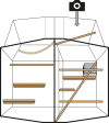
Fig. 6.. Amphetamine locomotion and stereotypy assessment.
Animals were segregated into the top half of their home cage (depicted) and a video camera placed on top of the transparent roof pointing down. Behavior was recorded for 45 min before a challenge (saline or amphetamine) and for 2.5 hours afterward.

References

    1. Heckers S., Konradi C., GABAergic mechanisms of hippocampal hyperactivity in schizophrenia. Schizophr Res. 167, 4–11 (2015). - PMC - PubMed
    1. Schobel S. A., Chaudhury N. H., Khan U. A., Paniagua B., Styner M. A., Asllani I., Inbar B. P., Corcoran C. M., Lieberman J. A., Moore H., Small S. A., Imaging patients with psychosis and a mouse model establishes a spreading pattern of hippocampal dysfunction and implicates glutamate as a driver. Neuron 78, 81–93 (2013). - PMC - PubMed
    1. Schobel S. A., Lewandowski N. M., Corcoran C. M., Moore H., Brown T., Malaspina D., Small S. A., Differential targeting of the CA1 subfield of the hippocampal formation by schizophrenia and related psychotic disorders. Arch. Gen. Psychiatry 66, 938–946 (2009). - PMC - PubMed
    1. Kaar S. J., Natesan S., McCutcheon R., Howes O. D., Antipsychotics: Mechanisms underlying clinical response and side-effects and novel treatment approaches based on pathophysiology. Neuropharmacology 172, 107704 (2020). - PubMed
    1. Shurman B., Horan W. P., Nuechterlein K. H., Schizophrenia patients demonstrate a distinctive pattern of decision-making impairment on the Iowa Gambling Task. Schizophr. Res. 72, 215–224 (2005). - PubMed

MeSH terms