. 2025 Apr 17;13(4):e010736.
doi: 10.1136/jitc-2024-010736.

Novel PD-1-targeted, activity-optimized IL-15 mutein SOT201 acting in cis provides antitumor activity superior to PD1-IL2v

Affiliations

Novel PD-1-targeted, activity-optimized IL-15 mutein SOT201 acting in cis provides antitumor activity superior to PD1-IL2v

Hana Matuskova et al. J Immunother Cancer. .

Abstract

Background: SOT201 and its murine surrogate mSOT201 are novel cis-acting immunocytokines consisting of a humanized/murinized/, Fc-silenced anti-programmed cell death protein 1 (PD-1) monoclonal antibody (mAb) fused to an attenuated human interleukin (IL)-15 and the IL-15Rα sushi+ domain. Murine mPD1-IL2v is a conjugate of a murinized, Fc silenced anti-PD-1 mAb bearing human IL-2 with abolished IL-2Rα binding. These immunocytokines spatiotemporally reinvigorate PD-1+ CD8+ tumor-infiltrating lymphocytes (TILs) via cis-activation and concomitantly activate the innate immunity via IL-2/15Rβγ signaling.

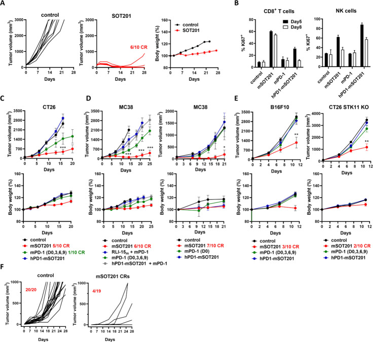
Methods: Human peripheral blood mononuclear cell and cell lines were used to evaluate cis/trans activity of SOT201. Anti-PD-1 mAb responsive (MC38, CT26) and resistant (B16F10, CT26 STK11 KO) mouse tumor models were used to determine the anticancer efficacy, and the underlying immune cell activity was analyzed via single-cell RNA sequencing and flow cytometry. The expansion of tumor antigen-specific CD8+ T cells by mSOT201 or mPD1-IL2v and memory CD8+ T-cell generation in vivo was determined by flow cytometry.

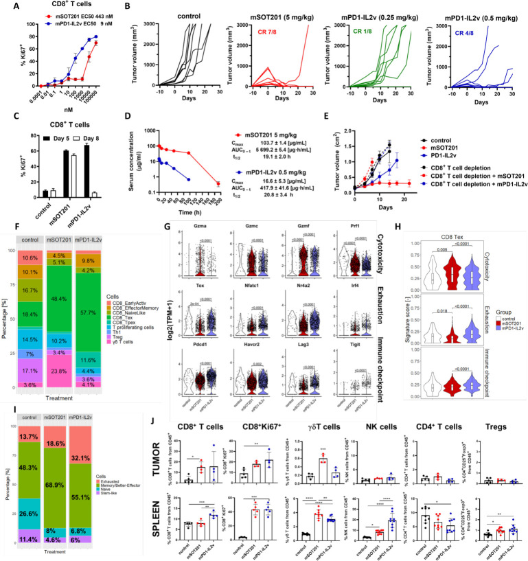
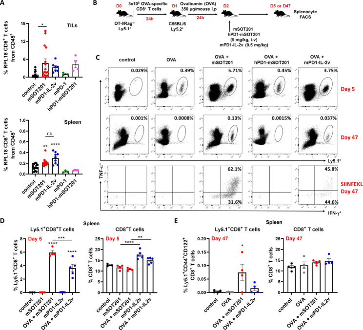
Results: SOT201 delivers attenuated IL-15 to PD-1+ T cells via cis-presentation, reinvigorates exhausted human T cells and induces higher interferon-γ production than pembrolizumab in vitro. mSOT201 administered as a single dose exhibits strong antitumor efficacy with several complete responses in all tested mouse tumor models. While mPD1-IL2v activates CD8+ T cells with a 50-fold higher potency than mSOT201 in vitro, mSOT201 more effectively reactivates effector exhausted CD8+ T cells (Tex), which demonstrate higher cytotoxicity, lower exhaustion and lower immune checkpoint transcriptional signatures in comparison to mPD1-IL2v in MC38 tumors in vivo. This can be correlated with a higher rate of complete responses in the MC38 tumor model following mSOT201 treatment when compared with mPD1-IL2v. mSOT201 increased the relative number of tumor antigen-specific CD8+ T cells, and unlike mPD1-IL2v stimulated greater expansion of adoptively transferred ovalbumin-primed CD8+ T cells simultaneously limiting the peripheral CD8+ T-cell sink, leading to the development of memory CD8+ T cells in vivo.

Conclusions: SOT201 represents a promising therapeutic candidate that preferentially targets PD-1+ TILs, delivering balanced cytokine activity for reviving CD8+ Tex cells in tumors. SOT201 is currently being evaluated in the Phase I clinical study VICTORIA-01 (NCT06163391) in patients with advanced metastatic cancer.

Keywords: Cytokine; Immune Checkpoint Inhibitor; Immunotherapy; T cell; Tumor microenvironment - TME.


Conflict of interest statement

Competing interests: All authors except VM, KB, MS, RM, MR and MK are employees of SOTIO Biotech a.s and DB is the CEO of Cytune Pharma.

Figures

Figure 1
Figure 1. Cis-acting SOT201 blocks PD-1/PD-L1 interactions, enhances IFN-γ production and reinvigorates partially exhausted human T cells in vitro. (A) Schematic representation of SOT201 molecule consisting of a humanized, Fc-silenced (L235E) IgG4 monoclonal antibody against PD-1 fused to one molecule of RLI-15 bearing N65A mutation. (B) Kit225 wt (expressing IL-2/15Rαβγ only) proliferation induced by SOT201, SOT201 wt (SOT201 without N65A mutation) and SOT101 (naked RLI-15, a complex of a human IL-15 linked to the IL-15Rα sushi+ domain). (C) Cis-acting SOT201 induces proliferation of Kit225 expressing PD-1+ and IL-2/15Rαβγ (Kit225 PD-1+). (D) Pembrolizumab blocks the SOT201-induced STAT-5 phosphorylation in Kit225-PD-1+. (E) No SOT201-induced STAT-5 phosphorylation in Kit225 wt in trans when co-incubated with Raji PD-1+ and lacking IL-2/15Rβγ. Representative of n=2–3. Schemes were created with BioRender.com. (F) SOT201-induced proliferation of PD-1+ and PD-1 hPBMC immune cell populations. Mean±SEM from 4 to 8 donors. (G) SOT201 blocks PD-1/PD-L1 interactions in vitro with IC50 of 1 nM (PD-L1 aAPC/CHO-K1 reporter cells). (H) SOT201 enhances IFN-γ production at 1 nM in mixed lymphocyte reaction (hPBMC) after 5 days in vitro. Mean±SEM of 12 donor pairs. (I) SOT201 reinvigorates partially exhausted human T cells in vitro. Means±SEM of 3 donors. hPBMC, human peripheral blood mononuclear cell; IFN, interferon; IL, interleukin; NK, natural killer; PD-1, programmed cell death 1; PD-L1, programmed cell death ligand 1; SEB, Staphylococcus aureus enterotoxin B; TIL, tumor-infiltrating lymphocyte; Treg, regulatory T cell.
Figure 2
Figure 2. SOT201 and mSOT201 induce strong antitumor efficacy in anti-PD-1 sensitive and resistant mouse tumor models via cis activation in vivo. (A) SOT201 (10 mg/kg, intravenous) or control treatment in hPD-1 transgenic mice bearing MC38-hPD-L1 tumors. (B) Splenic CD8+ and NK cell proliferation (Ki67+) in vivo 5 days after mSOT201, hPD1-mSOT201 (anti-human PD-1 mouse IgG1 clone RMP1-14 bearing D265A mutation in Fc region with attenuated RLI-15) and mPD-1 (clone RMP1-14) treatment at 5 mg/kg intravenously. Representative of n=2, two animals/group. SOT201 efficacy in anti-PD-1 sensitive mouse models. (C) CT26 (10 mg/kg all compounds), (D) MC38 (5 mg/kg mSOT201 or equimolar to the other compounds) and (E) in anti-PD-1 resistant mouse tumor models B16F10 or CT26 STK11 KO (10 mg/kg all compounds). Representative experiment, n=8–10 animals/group. (F) Rechallenge of naïve and SOT201-cured mice (after 100 days) with MC38 tumor cells (pool of experiments n=3). The treatment is day 0 if not stated otherwise. NK, natural killer; PD-1, programmed cell death 1.
Figure 3
Figure 3. mSOT201 expands cytotoxic CD8+ Tex with a better effector phenotype and γδ T cells in MC38 tumors. (A) UMAP plot of T-cell populations and γδ T cells in MC38 tumors 5 days after treatment with mSOT201, mPD-1 or hPD1-mSOT201 (5 mg/kg, intravenously). (B) Proportion of distinct T cell populations and γδ T cells. (C) Violin plots of selected genes in CD8+ Tex. (D) Violin gene signatures in CD8+ Tex. (E) The immune cells in tumors and spleen of MC38 tumor-bearing mice at day 5 post-treatment determined by flow cytometry. Mean±SEM. NK, natural killer; PD-1, programmed cell death 1; Tex, exhausted T cells; Treg, regulatory T cell; UMAP, Uniform Manifold Approximation and Projection.
Figure 4
Figure 4. mSOT201 induces better effector CD8+ Tex driving a superior antitumor response compared with mPD1-IL2v. (A) mSOT201 and mPD1-IL2v-induced splenic CD8+ T-cell proliferation after 5 days in vitro. (B) Antitumor efficacy in MC38 mouse tumor model. (C) Splenic CD8+ T-cell proliferation in vivo after 5 and 8 days after injection of mSOT201 (5 mg/kg intravenous) and mPD1-IL2v (0.25 mg/kg intravenous). (D) Pharmacokinetic profile in vivo. (E) Antitumor efficacy in MC38 mouse model±CD8+ T-cell depletion (F) T-cell populations (scRNA-seq) in MC38 tumors 5 days after treatment with mSOT201 (5 mg/kg intravenous) or mPD1-IL2v (0.5 mg/kg intravenous). (G) Violin plots of selected genes in CD8+ Tex. (H) Violin gene signature plots in CD8+ Tex. (I) Functional T cells (scRNA-seq) using an alternative clustering according to. (J) Flow cytometry phenotyping of MC38 tumors and spleen day 5 post-treatment. Means±SEM. NK, natural killer; PD-1, programmed cell death 1; scRNA-seq, single-cell RNA sequencing; Tex, exhausted T cells; Treg, regulatory T cell.
Figure 5
Figure 5. mSOT201 promotes memory formation in OVA-primed CD8+ T cells, while mPD1-IL2v expansion is limited by peripheral sink. (A) The RLP18-antigen specific CD8+ T cells from MC38 TILs and spleen 5 days after treatment. (B) Scheme of OT-I adoptive transfer. (C) Representative dotplots. (C, D) Quantification of OVA-specific CD8+ T cells (Ly5.1+) and endogenous CD8+ T-cell expansion on (C) day 5 and (D) day 47. All treatments were 5 mg/kg intravenous, except mPD1-IL2v was 0.5 mg/kg, intravenous. FACS, Fluorescence-Activated Cell Sorting; IL, interleukin; i.p, intraperitoneal; PD-1, programmed cell death 1; TILs, tumor-infiltrating lymphocytes.

References

    1. Waldmann TA. The shared and contrasting roles of IL2 and IL15 in the life and death of normal and neoplastic lymphocytes: implications for cancer therapy. Cancer Immunol Res. 2015;3:219–27. doi: 10.1158/2326-6066.CIR-15-0009. - DOI - PMC - PubMed
    1. Arneja A, Johnson H, Gabrovsek L, et al. Qualitatively different T cell phenotypic responses to IL-2 versus IL-15 are unified by identical dependences on receptor signal strength and duration. J Immunol. 2014;192:123–35. doi: 10.4049/jimmunol.1302291. - DOI - PMC - PubMed
    1. Meghnem D, Morisseau S, Frutoso M, et al. Cutting Edge: Differential Fine-Tuning of IL-2- and IL-15-Dependent Functions by Targeting Their Common IL-2/15Rβ/γc Receptor. J Immunol. 2017;198:4563–8. doi: 10.4049/jimmunol.1700046. - DOI - PubMed
    1. Stonier SW, Schluns KS. Trans-presentation: a novel mechanism regulating IL-15 delivery and responses. Immunol Lett. 2010;127:85–92. doi: 10.1016/j.imlet.2009.09.009. - DOI - PMC - PubMed
    1. Zhou Y, Husman T, Cen X, et al. Interleukin 15 in Cell-Based Cancer Immunotherapy. IJMS. 2022;23:7311. doi: 10.3390/ijms23137311. - DOI - PMC - PubMed

Associated data

LinkOut - more resources