. 2025 Apr 18;10(1):75.
doi: 10.1038/s41541-025-01123-y.

Intranasal spike and nucleoprotein fusion protein-based vaccine provides cross-protection and reduced transmission against SARS-CoV-2 variants

Affiliations

Intranasal spike and nucleoprotein fusion protein-based vaccine provides cross-protection and reduced transmission against SARS-CoV-2 variants

Zineb Lakhrif et al. NPJ Vaccines. .

Abstract

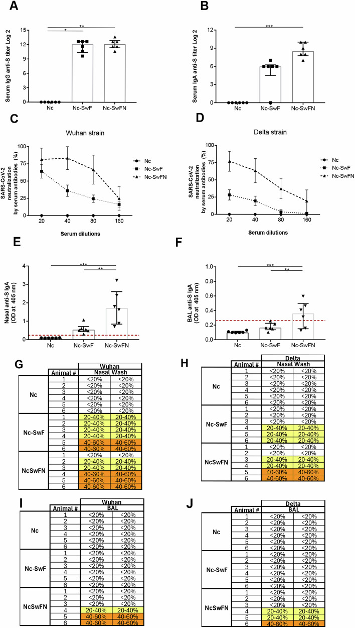
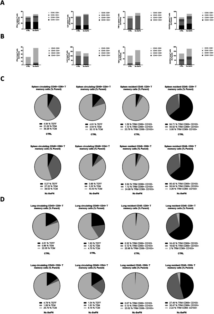
The effectiveness of intramuscular vaccines aimed at preventing severe COVID-19 remains limited due to waning immunity and the emergence of novel variants. Next-generation vaccines are needed for broader protection and blocking virus transmission. Here, we rationally designed an original nasal subunit vaccine composed of a fusion protein (SwFN) made of Wuhan spike and nucleoprotein combined with biocompatible mucosal nanocarriers (Nc). In mouse model, the nasal Nc-SwFN vaccine elicited multivalent serum and mucosal neutralizing antibodies. Robust spike and nucleoprotein cross-reactive immunity against variants was induced with a predominant phenotype of resident memory T cells in the lungs. Moreover, Nc-SwFN led to protective responses against Wuhan and Delta infection in relevant models with an absence of morbidity, mortality, and virus dissemination in the lungs and brain. Finally, Nc-SwFN drastically reduced host-to-host transmission. These promising results underscore the advantages of the nasal Nc-SwFN approach as a broad-spectrum vaccine candidate against current and emerging SARS-CoV-2 variants.


Conflict of interest statement

Competing interests: I.D.P., N.A., and M.E. are founders/equity holders in LovalTech. The remaining authors declare that the research was conducted without any commercial or financial relationships that could be construed as a potential conflict of interest.

Figures

Fig. 1
Fig. 1. Characterizations of SwFN protein and Nc-SwFN vaccine.
A Schematic representation of the heteromultimeric fusion protein. The two polypeptide chains constituting the fusion protein were presented at the far left, followed by the self-assembly thanks to the dimerization domain/ trimerization sequence block of the different chains. A schematic presentation of the isolated protein was mentioned on the right. B Schematic representation of the SwF heteromultimeric fusion protein without nucleoprotein. C Thermal analysis of the three proteins Sw, SwF, and SwFN. D exclusion-diffusion chromatography analysis of the SwF and SwFN fusion protein. E Sandwich ELISA shows the detection of the SwFN fusion protein by anti-N antibody after protein binding to the anti-S antibody. F Electrophoresis native PAGE of the SwFN fusion protein and vaccine complexations (Nc-SwF and Nc-SwFN). G Transmission electronic microscopy analysis of the SwFN (left), nanocarriers alone (medium), and the SwFN complexation with the nanocarriers (right). Arrowheads show examples of fusion proteins associated with the surface of the nanocarriers. Scale bar is 200 nm. H Vaccine uptake and I Internalization by the human nasal mucosa.
Fig. 2
Fig. 2. Humoral immune response against spike protein after SwFN and Nc-SwFN immunizations.
Female Balb/c mice were immunized twice at 3-week interval by intranasal route with nanocarriers alone (Nc) (n = 6 animals), SwFN (n = 6 animals), and Nc-SwFN complexation (n = 6 animals). Serum IgG and IgA antibodies were analyzed by specific anti-spike ELISA 7 days after the last immunization and presented respectively in (A, B). Their neutralization capacity was tested against Wuhan (analysis of serum pool) (C) and Delta (analysis of individual serum from 6 immunized mice) strains (D). Nasal and BAL anti-spike IgA antibodies were analyzed by specific anti-spike ELISA 7 days after the last immunization and presented respectively in (E, F). The neutralization capacity of the mucosal sample was tested against Wuhan (analysis of mucosal samples pooled from the 6 animals) (G, I) and Delta (analysis of mucosal samples individual) (H, J) strains, at the 1/5 dilution (column are experimental duplicates); nasal samples (G, H) and bronchoalveolar lavage samples (I, J). Data were analyzed by two-way ANOVA and Kruskal–Wallis tests (*p < 0.05, **p < 0.01, ****p < 0.0001).
Fig. 3
Fig. 3. Spike specific humoral immune response induced after Nc-SwF and Nc-SwFN immunizations.
Female Balb/c mice were immunized twice at 3-week interval by intranasal route with nanocarriers alone (Nc) (n = 6 animals), Nc-SwF (n = 6 animals) and Nc-SwFN complexation (n = 6 animals). IgG and IgA antibodies were analyzed by specific anti-spike ELISA 7 days after the last immunization. Serum anti-spike IgG and IgA were presented respectively in A, B with their neutralization capacity against Wuhan (C) and Delta strains (D). Nasal anti-spike IgA antibodies were showed in (E). BAL anti-spike IgA antibodies were showed in (F). The neutralization capacity of mucosal sample was tested against Wuhan (G, I) and Delta (H, J) strains, at the 1/5 dilution (column are experimental duplicates); nasal samples (G, H) and bronchoalveolar lavage samples (I, J). Data were analyzed by Kruskal–Wallis and one-way ANOVA tests (*p < 0.05, **p < 0.01, ***p < 0.001).
Fig. 4
Fig. 4. Systemic cellular immune response against nucleoprotein and spike SARS-CoV-2 variants after Nc-SwF and Nc-SwFN immunizations.
Female Balb/c mice were immunized twice at 3-week intervals by intranasal route with nanocarriers alone (Nc) (n = 6 animals), Nc-SwF (n = 6 animals) and Nc-SwFN complexation (n = 6 animals). Spleen and lung cells were stimulated by nucleoprotein and spike protein from SARS-CoV-2 Wuhan, Delta, and Omicron strains and IFN-γ (A, D, G, J, M, and P), IL-2 (B, E, H, K, N, and Q) and IL-17 (C, F, I, L, O, and R) cytokines were quantified by MACSPlex Cytokine kit after 72 hours. Data were analyzed by Kruskal–Wallis test (*p < 0.05, **p < 0.01).
Fig. 5
Fig. 5. Circulating and tissue-resident effector and memory T-cell subsets in spleens and lungs of Nc-SwFN immunized mice.
Female Balb/c mice were immunized twice at 3-week interval by intranasal route with Nc-SwFN complexation. Mice (n = 6 animals) were injected intravenously with anti-CD45-BV510 to distinguish circulatory and tissue-resident T cells. Intracellularly cytokine production (A, B) and T memory cell response (C, D) were analyzed in spleens and lungs, respectively. Gating strategies were showed in Supplementary figs. 4 and 5. Data were acquired on an MACSQuant®10 Analyzer (Miltenyi) and analyzed using FlowLogic software.
Fig. 6
Fig. 6. Evaluation of morbidity signs, survival mice, and virus presence after SARS-CoV-2 Delta infectious challenge.
Female K18-hACE2 mice were immunized twice at 3-week interval by intranasal route, with nanocarriers alone (Nc) (n = 6 animals), Nc-SwF (n = 6 animals) or with Nc-SwFN complexation (n = 6 animals). One or 4 weeks after the second immunization, mice were infected with Delta SARS-CoV-2 variant. Experimental timelines were indicated in (A). Mice body weight was taken daily post-infection (B). After infection, different clinicals signs were observed daily. Mice activity (C), respiratory distress (D), facies (E), and lordosis (F) were presented on day 8 post-infection. Mice survival was showed after infection with 1.3 × 105 TCID50 (G), and mice body weight and survival were showed after infection with 8 × 105 TCID50 (H, I) of Delta SARS-CoV-2 variant. To evaluate virus presence, histological sections of head, from infected mice with Delta SARS-CoV-2 variant (8 × 105 TCID50) since 7 or 8 days, were immunostained using SARS-CoV-2 N protein antibody (brown, scale bars: 1000 µm) (n = 6 heads for Nc and n = 3 heads for Nc-SwFN) (J). Survival data were analyzed by log-rank Mantel–Cox test (*p < 0.05, **p < 0.01).
Fig. 7
Fig. 7. Hamster protection associated with viral load abrogation in the lung and significant reduction in the nasal mucosa.
Male golden hamsters were immunized twice at three-week interval by intranasal route, with nanocarriers alone (Nc) (n = 4 animals) and Nc-SwFN complexation (n = 5 animals). One week after the second immunization, all hamsters were infected 5 × 104 TCID50 of SARS-CoV-2 Delta variant. Percentage of body weight change at day 2 post-infection compared to day 0 was presented in A. Lung tissues were collected at necropsy (day 2 post infection), and RNA was isolated for SARS-CoV-2 detection by qRT-PCR (B). Viral RNA relative loads compared to endogenous house-keeping control endogenous gene (2^(−deltaCt)) determined by qRT-PCR. A (2^(−deltaCt)) below 1 (dotted line) indicates no significant detection of viral RNA. For SARS-CoV-2 detection, lung tissues were processed for viral immunohistochemistry (IHC) using a mouse monoclonal anti-N antibody. Lung IHC scoring and representative images of lung sections IHC were showed in C, D, respectively. Ethmoid turbinates were collected at necropsy (day 2 post infection) and RNA was isolated for SARS-CoV-2 detection by qRT-PCR (E). Nasal swabs were collected on day 1 post-infection and infectious viral titers were determined by TCID50 (F). Schema of the hamster nasal cavity mucosa (Nt/Mt: Nasoturbinates and Maxilloturbinates, Et: Ethmoturbinates) (G). Representative images of nasal cavity mucosa viral IHC sections using a mouse monoclonal anti-N antibody (H). Higher magnification of Nt/mt and Et. The bars in the lung and nasal cavity figures represent 2 mm. Data were analyzed by a Mann–Whitney test (*p < 0.05).
Fig. 8
Fig. 8. Nc-SwFN vaccine protection against viral transmission.
Male golden hamsters were immunized twice at 3-week interval by intranasal route, with nanocarriers alone (Nc) (n = 5 animals) and Nc-SwFN complexation (n = 5 animals). One week after the second immunization, all hamsters were infected with 5 × 104 TCID50 of SARS-CoV-2 Delta variant. To evaluate the impact of vaccination on viral transmission post infection, naive hamsters were co-housed for 48 hours with the challenged animals at a ratio of 2 sentinels for 1 challenged animal (n = 10 animals co-housed with the n = 5 Nc immunized & challenged animals and n = 10 animals co-housed with the n = 5 Nc-SwFN immunized & challenged animals). The experimental protocol was schematized in A. Percentage of body weight change at day 3 post-infection, or day 3 post co-housing, compared to day 0 in the challenged animals (B) and the sentinel animals (F). Lung tissues and ethmoid turbinates were collected at necropsy (day 3 post-infection or day 3 post co-housing), and RNA of challenged (C, D) and sentinel (G, H) animals was isolated for SARS-CoV-2 detection by qRT-PCR. Viral RNA relative load compared to endogenous house-keeping control endogenous gene (2^(-deltaCt)) determined by qRT-PCR. A (2^(-deltaCt)) below 1 (dotted line) indicates no significant detection of viral RNA. Nasal swabs were collected on day 1 and day 2 post-infection or co-housing, and infectious viral titers of challenged (E) and sentinel (I) animals were determined by TCID50. Data were analyzed by a Mann–Whitney test (*p < 0.05; **p < 0.01).

References

    1. Belete, T. M. Review on up-to-date status of candidate vaccines for COVID-19 disease. IDRume 14, 151–161 (2021). - PMC - PubMed
    1. Hadj Hassine, I. Covid-19 vaccines and variants of concern: a review. Rev. Med. Virol.32, e2313 (2022). - PMC - PubMed
    1. A. M. Carabelli, et al. SARS-CoV-2 variant biology: immune escape, transmission and fitness. Nat. Rev. Microbiol.10.1038/s41579-022-00841-7 (2023). - PMC - PubMed
    1. Travis, C. R. As Plain as the nose on your face: the case for a nasal (Mucosal) route of vaccine administration for Covid-19 disease prevention. Front. Immunol.11, 591897 (2020). - PMC - PubMed
    1. Tiboni, M., Casettari, L. & Illum, L. Nasal vaccination against SARS-CoV-2: synergistic or alternative to intramuscular vaccines? Int. J. Pharm.603, 120686 (2021). - PMC - PubMed

LinkOut - more resources