. 2025 Apr 18;15(1):13464.
doi: 10.1038/s41598-025-92016-6.

Determining potential immunomodulatory drug efficacy in sepsis using ELISpot

Affiliations

Determining potential immunomodulatory drug efficacy in sepsis using ELISpot

Andrew H Walton et al. Sci Rep. .

Abstract

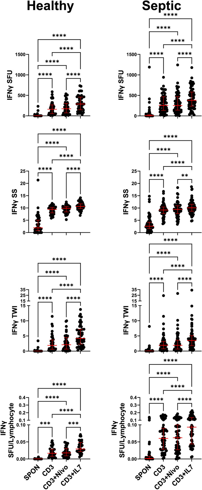
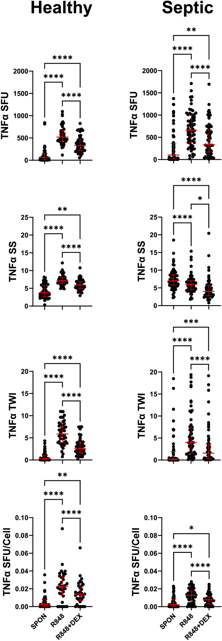
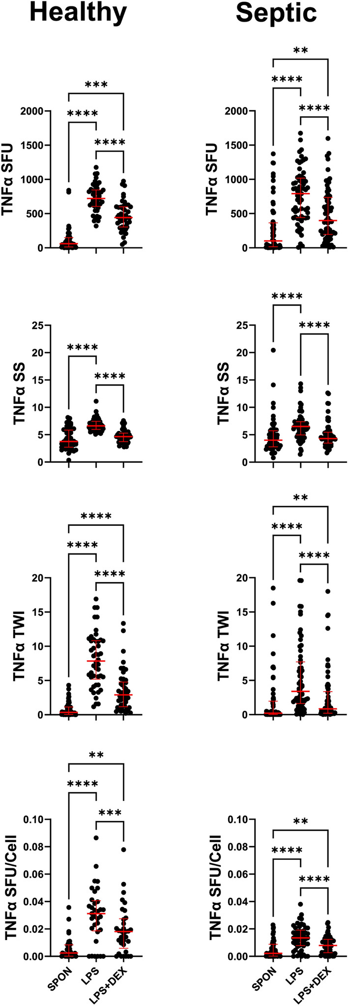
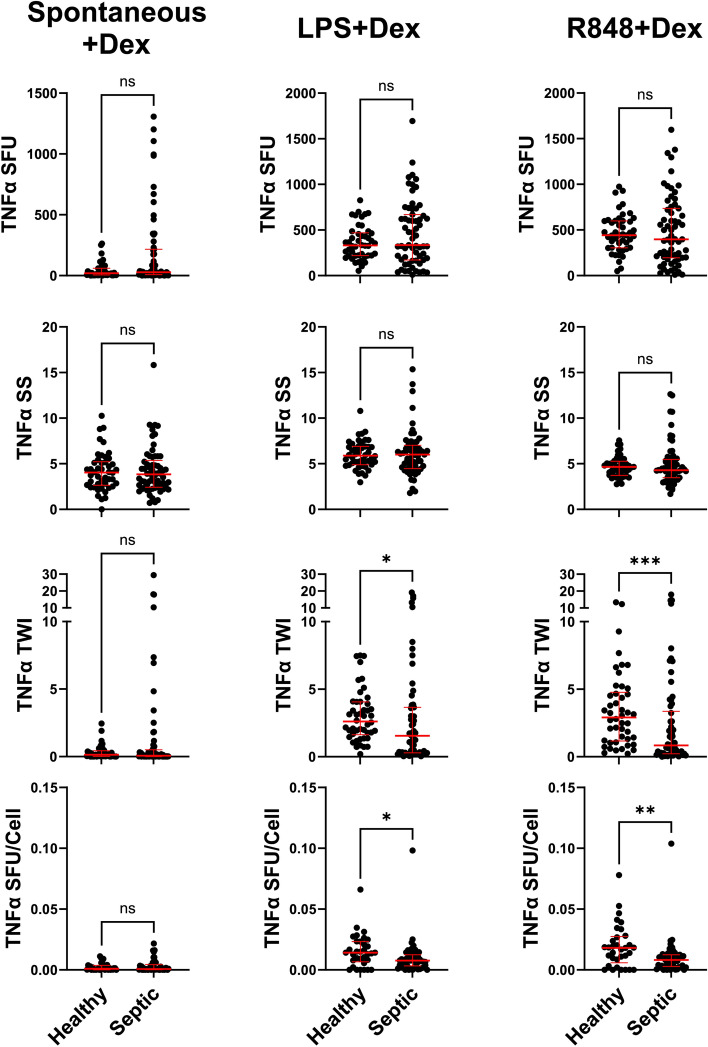
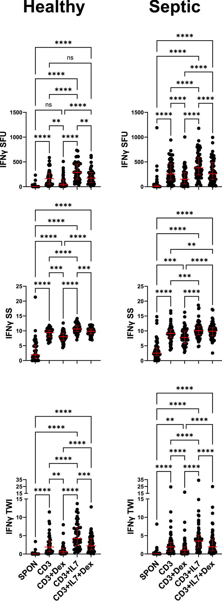
This study evaluated the ability of ELISpot to identify potential immuno-modulatory drug therapies in sepsis. ELISpot was performed ex vivo on whole blood from septic patients and healthy controls. Innate and adaptive immunity were evaluated by production of TNF-α and IFN-γ, respectively. Drug efficacy was determined by their effects to modulate the both the number of cytokine-producing cells and amount of cytokine produced per cell. The corticosteroid dexamethasone was evaluated for its ability to down modulate TNF-α and IFN-γ production. The TLR7/8 agonist resiquimod (R848) and T cell stimulants IL-7 and anti-PD-1 mAb were tested for their ability to enhance immunity. LPS and resiquimod increased total TNF-α production in septic patients by 1,549% and 1,829%, respectively. Conversely, dexamethasone diminished the responses to LPS or resiquimod by 75% and 61%, respectively. IL-7, but not anti-PD-1 mAb markedly increased IFN-γ production in both healthy subjects (121%) and septic patients (82%). Dexamethasone also reduced anti-CD3/CD28 mAb stimulated IFN-γ production by 69%; while IL-7 ameliorated dexamethasone-induced suppression. IL-7 significantly enhanced lymphocyte function in over 90% of septic patients. ELISpot can reveal host immune response patterns and the effects of drugs to selectively down- or up-regulate patient immunity. Furthermore, the ability of ELISpot to detect the effect of specific immuno-modulatory drugs to independently regulate the innate and adaptive host response could enable precision-based immune drug therapies in sepsis.

Keywords: Adaptive immunity; Anti-PD-1; Checkpoint inhibitors; Corticosteroids; IL-7; Innate immunity.


Conflict of interest statement

Declaration. Competing interests: MBM, and KER are members of Immune Functional Diagnostics, LLC and receive no direct financial compensation. Immune Functional Diagnostics, LLC is developing predictive metrics in critical illness and this technology (provisional patent application 63/521,817) is evaluated in this research. SCB, LLM, RSH, and the University of Florida may receive royalty income based on a technology developed by SCB and others and licensed by Washington University in St. Louis to IFDx LLC. That technology is evaluated in this research. CCC and the University of Cincinnati may receive royalty income based on a technology developed by CCC and others and licensed by Washington University in St. Louis to IFDx LLC. That technology is evaluated in this research.

Figures

Fig. 1
Fig. 1
Adjuvant effect on IFN-γ production in conjunction with anti-CD3/CD28 mAb stimulation. Samples were stimulated with anti-CD3/28 mAb with and without either anti-PD-1 mAb (Nivolumab; NIVO) or IL-7. Anti-CD/CD28 mAb (CD3) increased IFN-γ SFU (upper panels), SS (upper-middle panels) and TWI (lower-middle panels) in samples from both Healthy donors (left, n = 46) and Septic patients (right, n = 63) as compared to unstimulated spontaneous (SPON) production. NIVO did not increase CD3 induced IFN-γ SFU, SS, or TWI, while IL-7 did increase CD3 induced IFN-γ SFU, SS, and TWI. SFU (Top panels) were normalized to the number of lymphocytes plated in the ELISpot well (Bottom panels, n = 34 healthy and n = 59 septic). Lymphocyte number per well was determined by multiplying the draw specific ALC (K/cumm) by 1000 (converting K/cumm to cells/cumm) then by the volume of blood added to the well (5µL). Pairwise statistical relationships of SFU/Lymphocyte remain largely the same as SFU. Red bars represent Median with Interquartile Ranges (IQR) *p < 0.05, **p < 0.01, ***p < 0.001, ****p < 0.0001 as determined by Friedman Test with multiple comparisons.
Fig. 2
Fig. 2
Difference in relative IFN-γ response to IL-7 between healthy donors and septic patients. A. IL-sevenfold-changes were calculated for SFU, SS, and TWI for all samples; anti-CD3/CD28 mAb (CD3) + IL-7 values were divided by CD3 alone to yield fold-change. Samples from healthy donors displayed a higher responsiveness to IL-7 in the SFU (Left) and TWI (Right) measurements, based on fold-change, than did those from septic patients. B. Visualization of paired data points for CD3 alone or with IL-7 for samples from healthy donors (Left, n = 46) and septic patients (Right, n = 63). The bulk of samples show an increase of SFU and SS with IL-7 treatment. Red bars represent Median with IQR. *p < 0.05, **p < 0.01, ***p < 0.001, ****p < 0.0001 as determined by (A.) Mann–Whitney or (B.) Wilcoxon tests.
Fig. 3
Fig. 3
Septic patient samples produce greater IFN-γ SFU and SFU/Lymphocyte than Healthy donor samples. Comparison between Healthy donor (n = 46) and Septic patient (n = 63) whole blood samples shows septic blood samples have higher IFN-γ SFU (top) than healthy donor samples in response to anti-CD3/CD28 mAb (CD3) alone (left), CD3 + anti-PD-1 mAb (Nivolumab; NIVO) (middle), and CD3 + IL-7 (right). To account for high variability in cell counts among septic patients and between septic and healthy samples, SFU were normalized to number of lymphocytes plated in ELISpot wells (bottom, n = 34 healthy and n = 59 septic). When SFU were normalized to lymphocyte numbers, the differences between septic and healthy IFN-γ production became more apparent. As no significant differences were discerned between CD3 treated samples and CD3 + NIVO treated samples, the differences displayed in the “CD3 + NIVO” column between healthy and septic are a consequence of the greater responsiveness of septic samples to CD3 treatment. Red bars represent Median with IQR. *p < 0.05, **p < 0.01, ***p < 0.001, ****p < 0.0001 as determined by Mann–Whitney test.
Fig. 4
Fig. 4
Adjuvant effect of dexamethasone on TNF-α production in conjunction with resiquimod (R848) stimulation. Samples were stimulated with R848 with and without dexamethasone (DEX). R848 increased TNF-α SFU (upper panels), SS (upper-middle panels) and TWI (lower-middle panels) in samples from both Healthy donors (left, n = 46) and Septic patients (right, n = 63) as compared to unstimulated (spontaneous, SPON) production. DEX markedly abrogated R848 induced TNF-α production in SFU, SS, and TWI. SFU (Top panels) were normalized to the number of TNF-α producing cells (Cells) plated in the ELISpot well (Bottom panels, n = 34 healthy and n = 53 septic). The number of TNF-α producing cells per well was determined by multiplying the draw specific Absolute Neutrophil and Absolute Monocyte counts (ANC + AMC) in K/cumm by 1000 (converting K/cumm to cells/cumm) then by the volume of blood added to the well (5µL). Pairwise statistical relationships of SFU/Cell remain largely the same as SFU. Red bars represent Median with IQR. *p < 0.05, **p < 0.01, ***p < 0.001, ****p < 0.0001 as determined by Friedman Test with multiple comparisons.
Fig. 5
Fig. 5
Adjuvant effect of dexamethasone on TNF-α production in conjunction with Lipo-polysaccharide (LPS) stimulation. Samples were stimulated with LPS with and without dexamethasone (DEX). LPS increased TNF-α SFU (upper panels), SS (upper-middle panels) and TWI (lower-middle panels) in samples from both Healthy donors (left, n = 48) and Septic patients (right, n = 61) as compared to unstimulated (spontaneous, SPON) production. DEX markedly abrogated LPS induced TNF-α production in SFU, SS, and TWI. SFU (Top panels) were normalized to the number of TNF-α producing cells (Cells) plated in the ELISpot well (Bottom panels, n = 34 healthy and n = 53 septic). The number of TNF-α producing cells per well was determined by multiplying the draw specific Absolute Neutrophil and Absolute Monocyte counts (ANC + AMC) in K/cumm by 1000 (converting K/cumm to cells/cumm) then by the volume of blood added to the well (5µL). Pairwise statistical relationships of SFU/Cell remain largely the same as SFU. Red bars represent Median with IQR. *p < 0.05, **p < 0.01, ***p < 0.001, ****p < 0.0001 as determined by Friedman Test with multiple comparisons.
Fig. 6
Fig. 6
Comparison of responsiveness to dexamethasone between healthy donor and septic samples. Dexamethasone (DEX) was able to abrogate TNF-α production equally in healthy donor (n = 46) and septic patient (n = 63) samples for most parameters when looking at spontaneous TNF-α production (Left), LPS-induced TNF-α production (middle), or R848-induced TNF-α production (Right). DEX-based inhibition of LPS-induced TNF-α production was more pronounced in septic samples in the TWI measurement and R848-induced TNF-α production on the TWI measurement and on an SFU per cell basis (Bottom, n = 34 healthy and n = 53 septic) in septic patient than in healthy donor samples. Red bars represent Median with IQR. *p < 0.05, **p < 0.01, ***p < 0.001, ****p < 0.0001 as determined by Mann–Whitney test.
Fig. 7
Fig. 7
Effect of dexamethasone on IFN-γ production. IFN-γ SFU (Top), SS (Middle), and TWI (Bottom), are reduced when dexamethasone (DEX) is added to ELISpot. DEX abrogates anti-CD3/CD28 mAb (CD3) induced IFN-γ production. DEX-induced reduction of IFN-γ production is still present when co-cultured with anti-CD3/CD28 mAb and IL-7, though there is still an IL-7 effect present even with DEX co-treatment. Results were similar in healthy donor (n = 46) and septic patient (n = 63) samples. Red bars represent Median with IQR. *p < 0.05, **p < 0.01, ***p < 0.001, ****p < 0.0001 as determined by Friedman Test with multiple comparisons.

Similar articles

References

    1. Singer, M. et al. The third international consensus definitions for sepsis and septic shock (sepsis-3). JAMA315, 801–810. 10.1001/jama.2016.0287 (2016). - PMC - PubMed
    1. Bauer, M. et al. Mortality in sepsis and septic shock in Europe, North America and Australia between 2009 and 2019- results from a systematic review and meta-analysis. Crit. Care24, 239. 10.1186/s13054-020-02950-2 (2020). - PMC - PubMed
    1. Cecconi, M., Evans, L., Levy, M. & Rhodes, A. Sepsis and septic shock. Lancet392, 75–87. 10.1016/S0140-6736(18)30696-2 (2018). - PubMed
    1. Hotchkiss, R. S. et al. Sepsis and septic shock. Nat. Rev. Dis. Primers2, 16045. 10.1038/nrdp.2016.45 (2016). - PMC - PubMed
    1. Textoris, J. Immunity check should be performed for all patients with septic shock? Yes. Intensive Care Med.46, 503–505. 10.1007/s00134-019-05909-2 (2020). - PMC - PubMed

MeSH terms

LinkOut - more resources