. 2025 Apr 18;15(1):13377.
doi: 10.1038/s41598-025-96438-0.

Caerin 1.1 and 1.9 peptides induce acute caspase 3/GSDME-mediated pyroptosis in epithelial cancer cells

Affiliations

Caerin 1.1 and 1.9 peptides induce acute caspase 3/GSDME-mediated pyroptosis in epithelial cancer cells

Yuandong Luo et al. Sci Rep. .

Abstract

Caerin peptides exhibit a dual role in cancer treatment by directly killing cancer cells and modulating the tumour microenvironment to enhance anti-tumour immunity. This study investigates the mechanisms underlying caerin 1.1/1.9-induced acute cell death in epithelial cancer cells and explores their therapeutic potential. HeLa, A549, and Huh-7 cancer cell lines were treated with caerin 1.1/1.9 peptides. Morphological observations, flow cytometry, lactate dehydrogenase (LDH) release, and IL-18 secretion assays revealed the occurrence of pyroptosis following treatment. Specifically, a 1-h treatment with caerin 1.1/1.9 induced pyroptosis in HeLa, A549, and Huh-7 cells, characterised by cell swelling, membrane bubbling, and the release of IL-18 and LDH. Western blotting confirmed the upregulation of pyroptosis markers, including caspase-3, cleaved caspase-3, and GSDME-N fragments. These findings highlight the significant role of caerin peptides in inducing acute pyroptosis, a form of programmed cell death that enhances the immunogenicity of dying cancer cells, thus potentially improving the effectiveness of immunotherapies. This research underscores the therapeutic potential of caerin 1.1/1.9 peptides in cancer treatment, providing a foundation for developing new anti-cancer strategies that leverage both direct cytotoxic effects and immune modulation to achieve more effective and sustained anti-tumour responses.

Keywords: Caerin peptide; Caspase 3/GSDME signalling pathway; IL-18; Lactate dehydrogenase; Pyroptosis.


Conflict of interest statement

Declarations. Competing interests: The authors declare no competing interests.

Figures

Fig. 1
Fig. 1
The cell death induced by F1/F3 is not mediated by ferroptosis, necroptosis, or autophagy. (A) F1/F3 inhibits HeLa cell proliferation as assessed by MTT assay. (B) MTT assay determining the IC50 value of F1/F3 on HeLa cells. (C) Viability of HeLa cells after treatment with F1/F3 in combination with liproxstatin-1 (ferroptosis inhibitor). (D) Viability of HeLa cells after treatment with F1/F3 in combination with necrosulfonamide (necroptosis inhibitor). (E) Viability of HeLa cells after treatment with F1/F3 in combination with GSK-872 (necroptosis inhibitor). (F) Viability of HeLa cells after treatment with F1/F3 in combination with 3-MA (autophagy inhibitor). Different inhibitors could not restore the viability of F1/F3-treated HeLa cells, suggesting that the cell death induced by F1/F3 is not mediated by ferroptosis, necroptosis, or autophagy. All data are presented as the mean ± SEM. *P-value < 0.05, **P-value < 0.01, ***P-value < 0.001. Statistical significance was assessed using one-way ANOVA.
Fig. 2
Fig. 2
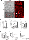
F1/F3 promotes pyroptosis of HeLa cells. (A) Real-time fluorescence images of HeLa cells treated with F1/F3 for 1 h. The HeLa cell membrane is stained with Dil. Scale bar: 50 μm. Black and white arrows point to membrane bubbles. (B) LDH release by HeLa cells after 1 h of F1/F3 treatment. (C) Levels of IL-1β secreted by HeLa cells after overnight treatment with different concentrations of F1/F3, measured by ELISA. (D) IL-18 secretion by HeLa cells treated overnight with P3, F1, F3, or F1/F3, measured by ELISA. (E) Levels of IL-18 secreted by HeLa cells after overnight treatment with different concentrations of F1/F3, measured by ELISA. (F) Levels of IL-18 secreted by HeLa cells at different time points during treatment with 10 µg/ml F1/F3, measured by ELISA. (G) Levels of IL-1β secreted by HeLa cells at different time points during treatment with 10 µg/ml F1/F3, measured by ELISA. All data are presented as the mean ± SEM. *P-value < 0.05, **P-value < 0.01, ***P-value < 0.001. Statistical significance was assessed using one-way ANOVA. These results are representative of two independent experiments.
Fig. 3
Fig. 3
F3/F3 promotes pyroptosis of A549 and Huh-7 cells. (A) Real-time fluorescence images of A549 cells treated with F1/F3 for 1 h, with the cell membrane stained with Dil and the nucleus stained with Hoechst 33,342. Scale bar: 50 μm. White arrows point to bubbles. (B) Real-time images of Huh-7 cells stimulated by F1/F3 for 1 h. Scale bar: 50 μm. Red arrows point to membrane bubbles. (C) MTT assay results showing the effect of 10 µg/ml F1/F3 on the proliferation of A549 cells. (D) MTT assay results showing the effect of 10 µg/ml F1/F3 on the proliferation of Huh-7 cells. (E) LDH release by A549 cells after 1 h of F1/F3 treatment. (F) MTT assay results showing the effect of 10 µg/ml F1/F3 on the proliferation of Huh-7 cells. (G) IL-18 secretion by A549 cells treated with F1/F3 at different time periods, measured by ELISA. (H) IL-1β secretion by A549 cells treated with F1/F3 at different time periods, measured by ELISA. All data are presented as the mean ± SEM. *P-value < 0.05, **P-value < 0.01, ***P-value < 0.001. Statistical significance was assessed using one-way ANOVA. These results are representative of two independent experiments.
Fig. 4
Fig. 4
F1/F3 mediates HeLa cell pyroptosis through the caspase-3/GSDME signalling pathway. (A)-(I): HeLa cells (1.0 × 106) were treated with F1/F3 (10 µg/ml), P3 or left untreated for 1 h, the expression of pyroptosis-related proteins was tested by Western blot. (A) GSDMD. (B) Caspase-1. (C) Caspase-4. (D) Caspase-5. (E) Cleaved caspase-4. (F) GSDMB. (G) Caspase-3. (H) Cleaved caspase-3. (I) Cleaved GSDME-N. (J) LDH release was detected by F1/F3, caspase inhibitor and caspase-3 inhibitor (Ac-DEVD-CHO) combined with F1/F3. These results come from a representative experiment in which two independent experiments were conducted. All data are presented as the mean ± SEM. *P-value < 0.05, **P-value < 0.01, ***P-value < 0.001. The statistical significance was assessed using one-way ANOVA.
Fig. 5
Fig. 5
F1/F3 treatment of HeLa cells for 1 h does not affect the expression of endogenous and exogenous apoptosis-related proteins. (A) Flow cytometric analysis and quantification of cell death by annexin V and propidium iodide (PI) staining. Early apoptosis is represented by Q3 (FITC + PI-) and late apoptosis, necrosis, or pyroptosis is represented by Q2 (FITC + PI+). (B)-(H): HeLa cells (1.0 × 106) were treated with F1/F3 (10 µg/ml), P3, or left untreated for 1 h, and protein expression levels of apoptotic-related proteins were measured by Western blot. (B) Caspase-8. (C) Cleaved caspase-8. (D) Bax. (E) Cytochrome c. (F) Apaf-1. (G) Caspase-7. (H) Cleaved caspase-7. These results come from a representative experiment in which two independent experiments were conducted. All data are presented as the mean ± SEM. *P-value < 0.05, **P-value < 0.01, ***P-value < 0.001. Statistical significance was assessed using one-way ANOVA.
Fig. 6
Fig. 6
A novel pathway for the induction of epithelial cancer cell death by caerin 1.1/1.9. One hour following caerin 1.1/1.9 treatment: HeLa cells activate caspase-3. Activated caspase-3 cleaves GSDME, resulting in membrane bubbling and the release of IL-18 and LDH. Following overnight treatment: HeLa cells upregulate the expression of Bax, downregulate Bcl-2, and show a decrease in mitochondrial membrane potential (MMP). This leads to the release of cytochrome c, which combines with Apaf-1 in the cytoplasm to form the apoptosome. The apoptosome activates caspase-9, which subsequently activates downstream caspase-3/7, resulting in HeLa cell apoptosis.

Similar articles

Cited by

References

    1. Bray, F. et al. Global cancer statistics 2022: GLOBOCAN estimates of incidence and mortality worldwide for 36 cancers in 185 countries. CA Cancer J. Clin.74, 229–263. 10.3322/caac.21834 (2024). - PubMed
    1. Cao, W., Chen, H. D., Yu, Y. W., Li, N. & Chen, W. Q. Changing profiles of cancer burden worldwide and in China: A secondary analysis of the global cancer statistics 2020. Chin. Med. J. (Engl)134, 783–791. 10.1097/cm9.0000000000001474 (2021). - PMC - PubMed
    1. Siegel, R. L., Giaquinto, A. N. & Jemal, A. Cancer statistics, 2024. CA Cancer J. Clin.74, 12–49. 10.3322/caac.21820 (2024). - PubMed
    1. Palanivelu, L., Liu, C. H. & Lin, L. T. Immunogenic cell death: the cornerstone of oncolytic viro-immunotherapy. Front. Immunol.13, 1038226. 10.3389/fimmu.2022.1038226 (2022). - PMC - PubMed
    1. Vivekanandhan, S. et al. Immunotherapies in rare cancers. Mol. Cancer22, 23. 10.1186/s12943-023-01720-2 (2023). - PMC - PubMed

LinkOut - more resources