

In vivo and in situ evaluation of innovative approaches in dentin hypersensitivity treatment
- PMID: 40251520
- PMCID: PMC12008923
- DOI: 10.1186/s12903-025-05865-y
In vivo and in situ evaluation of innovative approaches in dentin hypersensitivity treatment
Abstract
Background: Dentin hypersensitivity (DH) causes transient sharp pain from exposed dentinal tubules, adversely affecting oral health and quality of life. This study compared the efficacy of two innovative treatments against Sodium Fluoride Varnish in reducing DH and occluding dentinal tubules over eight weeks.
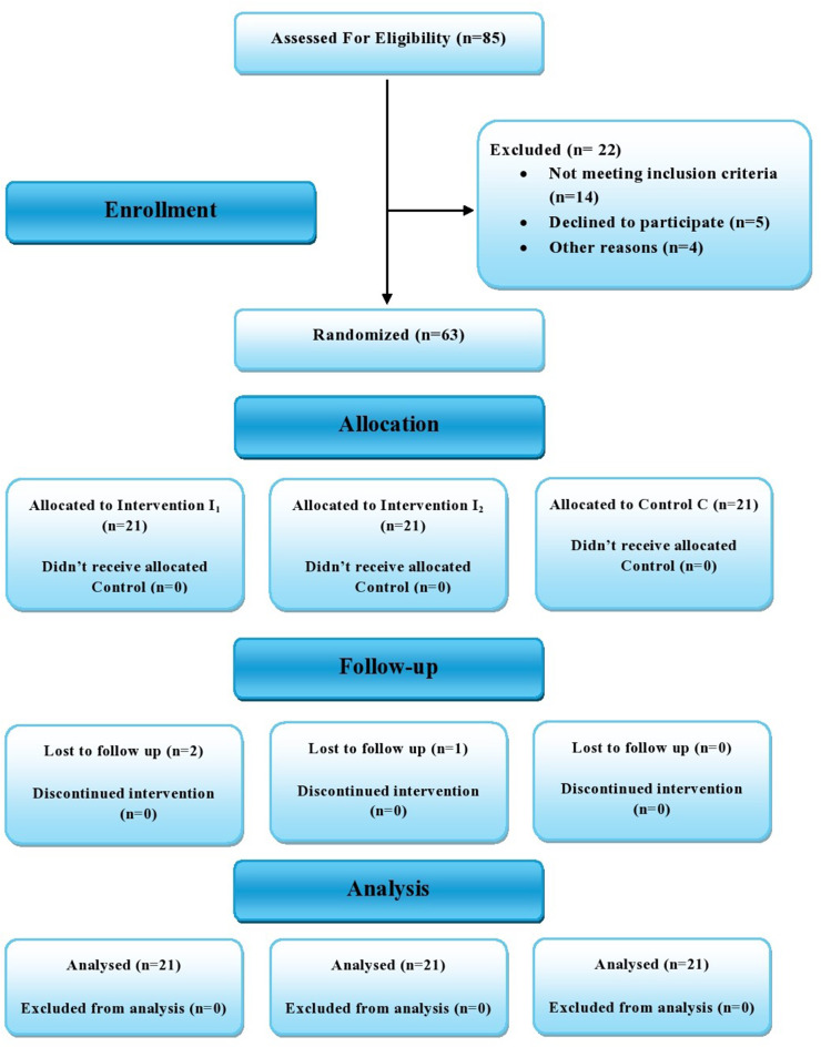
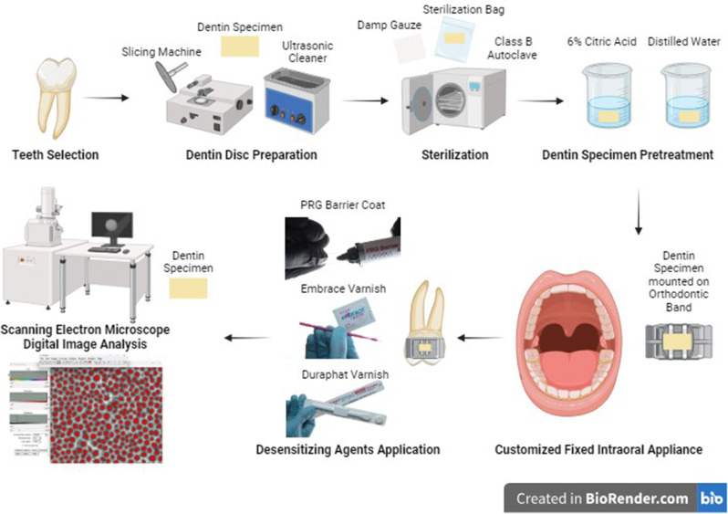
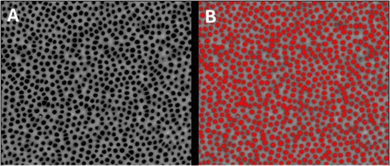
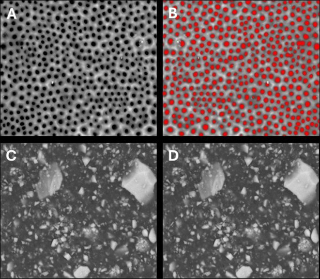
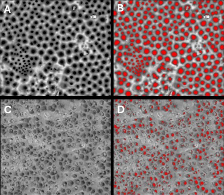
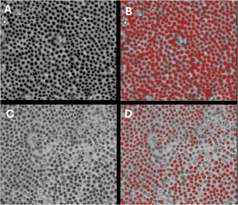
Methods: This randomized, triple-blind, three-parallel-arm clinical and in situ study included a total of 63 participants (age range: 26-46 years), each randomly assigned to one of three treatment groups: PRG Barrier Coat, Embrace varnish, or Duraphat varnish. The clinical trial assessed pain intensity was assessed using Visual Analog Scale (VAS) after tactile, evaporative, and thermal stimuli at baseline, 3 min, 2 weeks, 4 weeks, and 8 weeks. The in-situ phase evaluated dentinal tubules occlusion pre- and post-treatment using scanning electron microscopy (SEM) at 2000× magnification. Statistical Analysis was conducted using Kruskal-Wallis and Friedman tests for intergroup and intragroup comparisons, respectively, and Spearman's correlation for pain reduction-tubule occlusion relationship (p < 0.05).
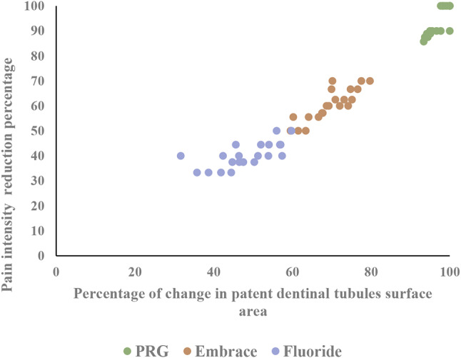
Results: PRG Barrier Coat achieved the highest efficacy with 94.9% pain reduction and 96.9% tubule occlusion. Embrace varnish showed moderate results with 64.3% pain reduction and 69.7% tubule occlusion, while Duraphat varnish provided limited performance with 45.4% pain reduction and 48.3% tubule occlusion. PRG Barrier Coat exhibited the most prolonged effects in reducing dentin hypersensitivity, aligning with its higher tubule occlusion. Embrace varnish demonstrated moderate performance, showing initial pain relief that was less sustained over time. Duraphat varnish provided the least reduction in pain and tubule occlusion, with effects that appeared transient.
Conclusions: This study demonstrated that PRG Barrier Coat and Embrace varnish effectively reduced pain intensity and promoted dentinal tubule occlusion, with PRG Barrier Coat showing the most sustained effects. These findings highlight the importance of dentinal tubule occlusion in DH management and suggest that treatment selection should consider both immediate pain relief and durability of therapeutic effects.
Trial registration: ClinicalTrials.gov (NCT04568473) on September 23, 2020.
Keywords: Dentin hypersensitivity; Dentinal tubules obliteration; Dentinal tubules occlusion; Duraphat; Embrace; Giomer; PRG barrier coat; Sodium fluoride; Xylitol-coated calcium phosphate.
© 2025. The Author(s).
Conflict of interest statement
Declarations. Human ethics and consent to participate declaration: This study received ethical approval from Research Ethics Committee (REC) of Faculty of Dentistry, Cairo University (Approval No. 19-9-20), in compliance with the Declaration of Helsinki and its subsequent amendments. The research was registered in ClinicalTrials.gov with unique identification number (NCT04568473) on September 23, 2020. Written informed consent was obtained from all participants included in the study. Consent to participate: Written informed consent was obtained in Arabic from all participants prior to their enrollment in the study, in accordance with the requirements of Research Ethics Committee (REC) at Faculty of Dentistry, Cairo University. Participants were informed that their names and personal information would remain confidential and would not be published. While every effort will be made to maintain anonymity, complete anonymity cannot be guaranteed. Consent for publication: Not Applicable. Competing interests: The authors declare no competing interests.
Figures
Similar articles
-
In vitro dentin permeability and tubule occlusion of experimental in-office desensitizing materials.Clin Oral Investig. 2023 Mar;27(3):1265-1276. doi: 10.1007/s00784-022-04760-y. Epub 2022 Oct 28. Clin Oral Investig. 2023. PMID: 36305964
-
Clinical efficiency of a natural resin fluoride varnish (Shellac F) in reducing dentin hypersensitivity.J Oral Rehabil. 2009 Feb;36(2):124-31. doi: 10.1111/j.1365-2842.2008.01907.x. J Oral Rehabil. 2009. PMID: 19522897 Clinical Trial.
-
Novel Management of Hypersensitive Dentin Using Propolis-based Herbal Desensitizing Agents: An In Vitro Scanning Electron Microscopic Study.J Contemp Dent Pract. 2021 Sep 1;22(9):1030-1034. J Contemp Dent Pract. 2021. PMID: 35000948
-
Treating hypersensitivity with fluoride varnishes.Compend Contin Educ Dent. 1998 Nov;19(11):1088-90, 1092, 1094 passim. Compend Contin Educ Dent. 1998. PMID: 10202463 Review.
-
Effect of desensitizing agents on dentin hypersensitivity after non-surgical periodontal therapy: A systematic review and meta-analysis.J Dent. 2020 Dec;103:103498. doi: 10.1016/j.jdent.2020.103498. Epub 2020 Oct 15. J Dent. 2020. PMID: 33069772
References
-
- Holland GR, Narhi MN, Addy M, Gangarosa L, Orchardson R. Guidelines for the design and conduct of clinical trials on dentine hypersensitivity. J Clin Periodontol. 1997;24(11):808–13. 10.1111/j.1600-051X.1997.tb01194.x. - PubMed
-
- Douglas-de-Oliveira DW, Vitor GP, Silveira JO, Martins CC, Costa FO, Cota LOM. Effect of dentin hypersensitivity treatment on oral health-related quality of life—A systematic review and meta-analysis. J Dent. 2018;71:1–8. 10.1016/j.jdent.2017.12.007. - PubMed
-
- Marto CM, Baptista Paula A, Nunes T, Pimenta M, Abrantes AM, Pires AS, et al. Evaluation of the efficacy of dentin hypersensitivity treatments—A systematic review and follow-up analysis. J Oral Rehabil. 2019;46(10):952–90. 10.1111/joor.12842. - PubMed
-
- Mosquim V, Caracho RA, Zabeu GS, Condi LS, Foratori-Junior GA, Borges AFS, et al. Resin-based materials to control human dentin permeability under erosive conditions in vitro: A hydraulic conductance, confocal microscopy, and FTIR study. Dent Mater. 2022;38(10):1669–78. 10.1016/j.dental.2022.08.012. - PubMed
Publication types
MeSH terms
Substances
Associated data
LinkOut - more resources
Full Text Sources
Medical
