

Evaluation of submandibular fossa depth and mandibular canal relationship with cone-beam computed tomography
- PMID: 40251541
- PMCID: PMC12008885
- DOI: 10.1186/s12903-025-05686-z
Evaluation of submandibular fossa depth and mandibular canal relationship with cone-beam computed tomography
Abstract
Background: Before performing surgical procedures in the posterior mandible, which contains vital structures, it is essential to have detailed knowledge of the morphology, depth, types, and variations of the submandibular fossa (SMF), as well as the precise course of the mandibular canal (MC) to avoid a possible perforation. The aim of this study was to evaluate the relationship between the depth and types of SMF, and the MC position using cone beam computed tomography (CBCT) images in a sample of Turkish individuals.
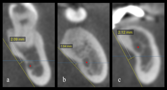
Methods: The depth and type of SMF, and relative position of the MC to the deepest point of the SMF (classified as inferior, parallel, or superior), were retrospectively evaluated on cross-sectional CBCT images. The Wilcoxon Signed Rank Test, Mann-Whitney U test, Spearman correlation coefficient, Kruskal-Wallis H test, and Chi-square test were used to analysis of the variables. A p value of less than 0.05 was accepted as significant.
Results: The submandibular fossa depth (SMFD) was measured on the left and right sides of CBCT images of 610 patients (358 female, 252 male). A significant difference was observed between the left and right sides for mean SMFD (1.93 ± 0.65 and 1.98 ± 0.67, respectively) (p = 0.001). The mean SMFD values were found to be significantly higher in males than in females on both sides (p < 0.001). The position of the MC in relation to the deepest point of the SMF was most frequently observed to be inferior. The lowest mean SMFD value was observed in the parallel position on the left and right sides (1.79 ± 0.56 and 1.77 ± 0.53, respectively).
Conclusions: The anatomical data obtained from this study contribute to the existing literature on SMFD and its relationship with the MC. It is recommended that these anatomical structures be evaluated radiologically before surgical procedures to prevent complications.
Keywords: CBCT; Mandibular Canal; Radiographic examination; Submandibular fossa.
© 2025. The Author(s).
Conflict of interest statement
Declarations. Ethics approval and consent to participate: This research protocol was approved by the Clinical Research Ethical Committee of Ordu University with the assignment protocol number 2023/79. All research procedures were conducted in accordance with the relevant guidelines and regulations set forth in the Declaration of Helsinki. This study was conducted retrospectively and did not contain any personally identifiable information. Informed consent to anonymized data release was obtained from all patients included in this study. Consent for publication: Not applicable. Competing interests: The authors declare no competing interests.
Figures


References
-
- Parnia F, Fard EM, Mahboub F, Hafezeqoran A, Gavgani FE. Tomographic volume evaluation of submandibular fossa in patients requiring dental implants. Oral Surg Oral Med Oral Pathol Oral Radiol Endod. 2010;109:e32–6. - PubMed
-
- de Souza LA, Souza Picorelli Assis NM, Ribeiro RA, Pires Carvalho AC, Devito KL. Assessment of mandibular posterior regional landmarks using cone-beam computed tomography in dental implant surgery. Ann Anat. 2016;205:53–9. - PubMed
-
- Herranz-Aparicio J, Marques J, Almendros-Marqués N, Gay-Escoda C. Retrospective study of the bone morphology in the posterior mandibular region. Evaluation of the prevalence and the degree of lingual concavity and their possible complications. Med Oral Patol Oral Cir Bucal. 2016;21:e731–6. - PMC - PubMed
-
- Rusu MC, Stoenescu MD, Butucescu M, Săndulescu M. The sand watch mandible. Folia Morphol (Warsz). 2023;82(2):424–8. - PubMed
-
- Tomljenovic B, Herrmann S, Filippi A, Kühl S. Life-threatening hemorrhage associated with dental implant surgery: a review of the literature. Clin Oral Implants Res. 2016;27:1079–84. - PubMed
MeSH terms
LinkOut - more resources
Full Text Sources
