. 2025 Apr 19;20(1):395.
doi: 10.1186/s13018-025-05809-w.

The effects of different exercise training protocols on mitochondrial dynamics in skeletal and cardiac muscles of Wistar rats

Affiliations

The effects of different exercise training protocols on mitochondrial dynamics in skeletal and cardiac muscles of Wistar rats

Amir Hossein Haghighi et al. J Orthop Surg Res. .

Abstract

Background: Mitochondrial fission and fusion both contribute to maintaining mitochondrial function and optimizing bioenergetic capacity.

Objective: The aim of this study was to compare the effect of aerobic and resistance training on mitochondrial fission and fusion markers in skeletal and cardiac muscles of Wistar rats.

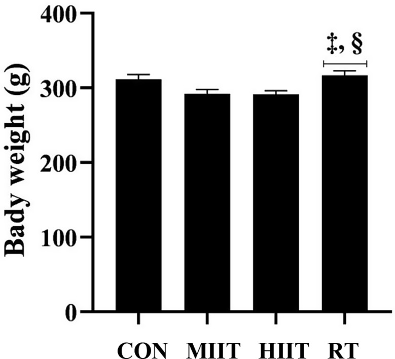
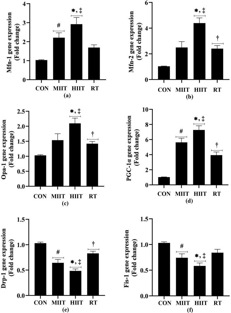
Method: 24 male Wistar rats were randomly divided into four groups of moderate-intensity interval training (MIIT), high-intensity interval training (HIIT), resistance training (RT) and control (CON). The MIIT and HIIT groups performed treadmill exercises with an intensity of 60-65% and 80-85% of the maximum speed, respectively, while the RT group performed resistance training with an intensity of 30-60% of the rat's body weight for 8 weeks. The soleus (SOL), extensor digitorum longus (EDL) and left ventricular tissues were used to evaluate markers of mitochondrial fission and fusion PGC-1α (fusion/fission), Opa-1 (fusion), Fis-1 (fission), Drp-1 (fission), Mfn-1 and Mfn-2 (fusion) genes expression.

Results: In all three tissues, a significant increase in some mitochondrial fusion markers was observed after 8 weeks of training (p = < 0.0001-0.0452). Furthermore, a significant decrease in cardiac mitochondrial fission markers was observed in all three groups (p = < 0.0001-0.0156). This reduction in some markers was evident in the SOL tissue of the HIIT group (p < 0.0001 for Drp-1 and p = 0.0007 for Fis-1) and in the EDL tissue of the RT group (p = 0.0005 for Fis-1 and p = 0.0012 for Drp-1). The mitochondrial fission/fusion markers in the heart (p = 0.0007-0.0449) and SOL (p = 0.0050-0.0258) tissues of the HIIT group had more changes than the RT group, while the mitochondrial fission markers in the EDL tissue of the RT group had a lower level than the HIIT (p = 0.0087 for Drp-1) and MIIT (p = 0.0130 for Fis-1 and p = 0.0010 for Drp-1) groups.

Conclusion: Our study demonstrated that HIIT, through better regulation of mitochondrial fusion and fission than RT, improves mitochondrial dynamics in cardiac and SOL tissues.

Keywords: Exercise; Mitochondrial dynamics; Muscles; Myocardium; Rats.


Conflict of interest statement

Declarations. Ethical approval: The animal study protocol was approved by the Institutional Review Board of Hakim Sabzevari University (protocol code IR.HSU.AEC.1401.017 on January 21th, 2023). Consent for publication: Not applicable in this section. Competing interests: The authors declare no competing interests.

Figures

Fig. 1
Fig. 1
Final weight of rats in study groups (The data presented as mean ± SEM). ‡: significant differences between HIIT and RT groups, §: significant differences between MIIT and RT groups. CON: control, MIIT: moderate-intensity interval training, HIIT: high-intensity interval training, RT: resistance training
Fig. 2
Fig. 2
The effect of training on the gene expression of mitochondrial indices in SOL muscle (The data presented as mean ± SEM). (a) Mfn-1 gene expression, (b) Mfn-2 gene expression, (c) Opa-1 gene expression, (d) PGC-1α gene expression, (e) Drp-1 gene expression, (f) Fis-1 gene expression. *significant differences between HIIT and CON groups, #: significant differences between MIIT and CON groups, †: significant differences between RT and CON groups, ‡: significant differences between HIIT and RT groups, £: significant differences between HIIT and MIIT groups. CON: control, MIIT: moderate-intensity interval training, HIIT: high-intensity interval training, RT: resistance training, SOL: soleus, Mfn-1: mitofusin-1, Mfn-2: mitofusin-2, Opa-1: optic atrophy-1, PGC-1α: peroxisome proliferator-activated receptor gamma co-activator 1 alpha, Drp-1: dynamin-related protein-1, Fis-1: mitochondrial fission-1
Fig. 3
Fig. 3
The effect of training on the gene expression of mitochondrial indices in EDL muscle (The data presented as mean ± SEM). (a) Mfn-1 gene expression, (b) Mfn-2 gene expression, (c) Opa-1 gene expression, (d) PGC-1α gene expression, (e) Drp-1 gene expression, (f) Fis-1 gene expression. *significant differences between HIIT and CON groups, #: significant differences between MIIT and CON groups, †: significant differences between RT and CON groups, ‡: significant differences between HIIT and RT groups, §: significant differences between MIIT and RT groups. CON: control, MIIT: moderate-intensity interval training, HIIT: high-intensity interval training, RT: resistance training, EDL: extensor digitorum longus, Mfn-1: mitofusin-1, Mfn-2: mitofusin-2, Opa-1: optic atrophy-1, PGC-1α: peroxisome proliferator-activated receptor gamma co-activator 1 alpha, Drp-1: dynamin-related protein-1, Fis-1: mitochondrial fission-1
Fig. 4
Fig. 4
The effect of training on the gene expression of mitochondrial indices in heart muscle (The data presented as mean ± SEM). (a) Mfn-1 gene expression, (b) Mfn-2 gene expression, (c) Opa-1 gene expression, (d) PGC-1α gene expression, (e) Drp-1 gene expression, (f) Fis-1 gene expression. *significant differences between HIIT and CON groups, #: significant differences between MIIT and CON groups, †: significant differences between RT and CON groups, ‡: significant differences between HIIT and RT groups. CON: control, MIIT: moderate-intensity interval training, HIIT: high-intensity interval training, RT: resistance training, Mfn-1: mitofusin-1, Mfn-2: mitofusin-2, Opa-1: optic atrophy-1, PGC-1α: peroxisome proliferator-activated receptor gamma co-activator 1 alpha, Drp-1: dynamin-related protein-1, Fis-1: mitochondrial fission-1

References

    1. Chen W, Zhao H, Li Y. Mitochondrial dynamics in health and disease: mechanisms and potential targets. Signal Transduct Target Ther. 2023;8:333. - PMC - PubMed
    1. Vezza T, Díaz-Pozo P, Canet F, de Marañón AM, Abad-Jiménez Z, García-Gargallo C, et al. The role of mitochondrial dynamic dysfunction in Age-Associated type 2 diabetes. World J Mens Health. 2022;40:399–411. - PMC - PubMed
    1. Li Y-J, Cao Y-L, Feng J-X, Qi Y, Meng S, Yang J-F, et al. Structural insights of human mitofusin-2 into mitochondrial fusion and CMT2A onset. Nat Commun. 2019;10:4914. - PMC - PubMed
    1. Alghamdi A. A detailed review of Pharmacology of MFN1 (mitofusion-1)-mediated mitochondrial dynamics: implications for cellular health and diseases. Saudi Pharm J. 2024;32:102012. - PMC - PubMed
    1. You W, Knoops K, Berendschot TTJM, Benedikter BJ, Webers CAB, Reutelingsperger CPM, et al. PGC-1a mediated mitochondrial biogenesis promotes recovery and survival of neuronal cells from cellular degeneration. Cell Death Discov. 2024;10:180. - PMC - PubMed

MeSH terms

Substances

LinkOut - more resources