

Generation and purification of iPSC-derived cardiomyocytes for clinical applications
- PMID: 40251664
- PMCID: PMC12008852
- DOI: 10.1186/s13287-025-04319-0
Generation and purification of iPSC-derived cardiomyocytes for clinical applications
Abstract
Background: Over the past decade, the field of cell therapy has rapidly expanded with the aim to replace and repair damaged cells and/or tissue. Depending on the disease many different cell types can be used as part of such a therapy. Here we focused on the potential treatment of myocardial infarction, where currently available treatment options are not able to regenerate the loss of healthy heart tissue.
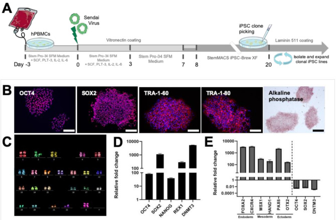
Method: We generated good manufacturing practice (GMP)-compatible cardiomyocytes (iCMs) from transgene- and xenofree induced pluripotent stem cells (iPSCs) that can be seamless adapted for clinical applications. Further protocols were established for replating and freezing/thawing iCMs under xenofree conditions.
Results: iCMs showed a cardiac phenotype, with the expression of specific cardiac markers and absence of pluripotency markers at RNA and protein level. To ensure a pure iCMs population for in vivo applications, we minimized risks of iPSC contamination using RNA-switch technology to ensure safety.
Conclusion: We describe the generation and further processing of xeno- and transgene-free iCMs. The use of GMP-compliant differentiation protocols ab initio facilitates the clinical translation of this project in later stages.
Keywords: Cardiomyocytes; Cell therapy; Clinical translation; Induced pluripotent stem cells.
© 2025. The Author(s).
Conflict of interest statement
Declarations. Ethical approval and consent to participate: Human peripheral blood was collected with written informed consent according to the permission from the cantonal ethics commission of Zurich, Switzerland [KEK-ZH-2014-0430] entitled “Periphere mononukleäre Blutzellen als Quelle für Tissue Engineering in der Regenerativen Medizin” (Amendmend 05.01.2015). The veterinary office of the Canton Zurich, Switzerland approved all animal experiments (ZH174/2020) entitled “Safety assessment of induced pluripotent stem cell-derived cardiomyocytes” (approved 19.02.2021). Consent for publication: All authors agreed to publication. Competing interests: S.P.H. is a shareholder at Xeltis BV and LifeMatrix Technologies AG. M.Y.E. is a shareholder at LifeMatrix Technologies AG. H.S. is the investigator of a record listed on a patent application (PCT/JP2017/023513, filed by Kyoto University on 27 June 2017) related to the design of the RNA-ON switch. H.S. is listed on a patent application (Japanese patent application no. 2021-177971) related to the cell purification. H.S. own shares of aceRNA Technologies Ltd, where H.S. is an outside director. The authors declare that they have no other competing interests.
Figures


Similar articles
-
Induced Pluripotent Stem Cell-Derived Cardiomyocytes: From Regulatory Status to Clinical Translation.Tissue Eng Part B Rev. 2024 Aug;30(4):436-447. doi: 10.1089/ten.TEB.2023.0080. Epub 2024 Feb 19. Tissue Eng Part B Rev. 2024. PMID: 38149607 Review.
-
The Future of Direct Cardiac Reprogramming: Any GMT Cocktail Variety?Int J Mol Sci. 2020 Oct 26;21(21):7950. doi: 10.3390/ijms21217950. Int J Mol Sci. 2020. PMID: 33114756 Free PMC article. Review.
-
Long-Term Stability and Differentiation Potential of Cryopreserved cGMP-Compliant Human Induced Pluripotent Stem Cells.Int J Mol Sci. 2019 Dec 23;21(1):108. doi: 10.3390/ijms21010108. Int J Mol Sci. 2019. PMID: 31877913 Free PMC article.
-
Efficient Cardiac Differentiation of Human Amniotic Fluid-Derived Stem Cells into Induced Pluripotent Stem Cells and Their Potential Immune Privilege.Int J Mol Sci. 2020 Mar 29;21(7):2359. doi: 10.3390/ijms21072359. Int J Mol Sci. 2020. PMID: 32235313 Free PMC article.
-
Xeno-free induced pluripotent stem cell-derived neural progenitor cells for in vivo applications.J Transl Med. 2022 Sep 16;20(1):421. doi: 10.1186/s12967-022-03610-5. J Transl Med. 2022. PMID: 36114512 Free PMC article.
References
-
- Tsao CW, et al. Heart disease and stroke statistics—2023 update: a report from the American heart association. Circulation. 2023;147(8):e93–621. - PubMed
MeSH terms
Grants and funding
LinkOut - more resources
Full Text Sources
Miscellaneous
