Review
. 2025 Mar-Apr;177(2):e70230.
doi: 10.1111/ppl.70230.

Lipid polymorphism of plant thylakoid membranes. The dynamic exchange model - facts and hypotheses

Affiliations
Review

Lipid polymorphism of plant thylakoid membranes. The dynamic exchange model - facts and hypotheses

Győző Garab et al. Physiol Plant. 2025 Mar-Apr.

Abstract

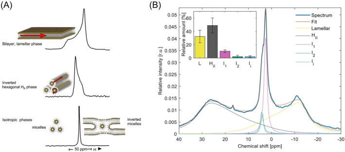
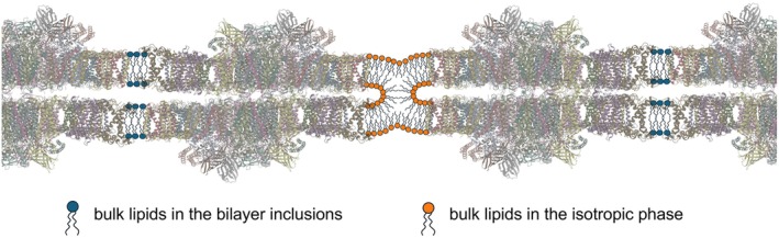
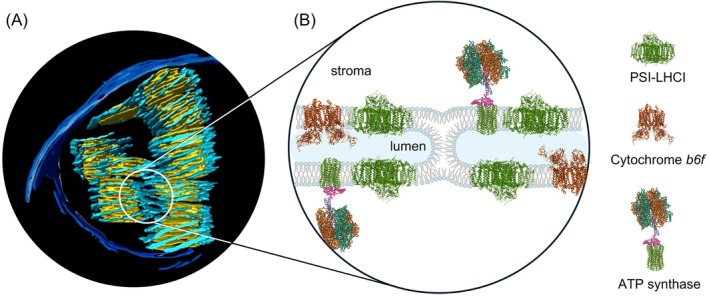
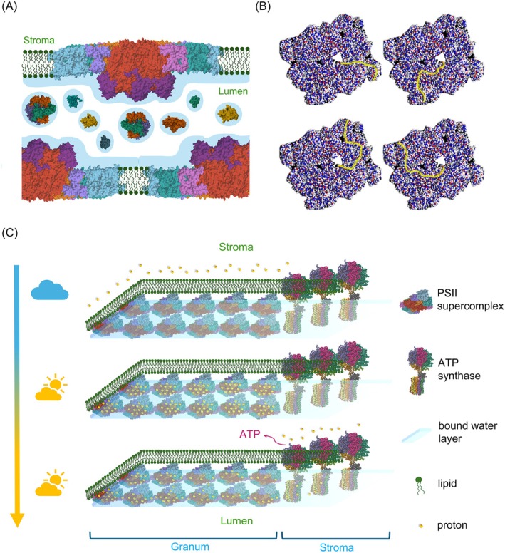
The light reactions of oxygenic photosynthesis are performed by protein complexes embedded in the lipid bilayer of thylakoid membranes (TMs). Bilayers provide optimal conditions for the build-up of the proton motive force (pmf) and ATP synthesis. However, functional plant TMs, besides the bilayer, contain an inverted hexagonal (HII) phase and isotropic phases, a lipid polymorphism due to their major, non-bilayer lipid species, monogalactosyldiacylglycerol (MGDG). The lipid phase behavior of TMs is explained within the framework of the Dynamic Exchange Model (DEM), an extension of the fluid-mosaic model. DEM portrays the bilayer phase as inclusions between photosynthetic supercomplexes - characterized by compromised membrane impermeability and restricted sizes inflicted by the segregation propensity of lipid molecules, safe-guarding the high protein density of TMs. Isotropic phases mediate membrane fusions and are associated with the lumenal lipocalin-like enzyme, violaxanthin de-epoxidase. Stromal-side proteins surrounded by lipids give rise to the HII phase. These features instigate experimentally testable hypotheses: (i) non-bilayer phases mediate functional sub-compartmentalization of plant chloroplasts - a quasi-autonomous energization and ATP synthesis of each granum-stroma TM assembly; and (ii) the generation and utilization of pmf depend on hydrated protein networks and proton-conducting pathways along membrane surfaces - rather than on strict impermeability of the bilayer.


Figures

FIGURE 1
FIGURE 1
Fingerprinting the lipid phase behavior of TMs by 31 P‐NMR spectroscopy. (A) Bilayer and non‐bilayer lipid assemblies, indicating also the characteristic motions of lipid molecules (red arrows), and the corresponding 31P‐NMR spectra (based on Cullis and de Kruijff 1979). (B) 31P‐NMR spectrum of freshly isolated spinach thylakoid membranes (thick dark blue trace) and the fitted spectrum (orange curve) obtained after deconvolution of the spectral components. The area‐normalized spectrum represents the average obtained from 52 independent experiments performed at 5°C. The average Chl content of these samples was 8.3 ± 3.1 mg ml−1. Individual contributions of the lamellar (L, yellow), inverted hexagonal (HII, grey), and isotropic (I1, I2, and Ii, pink, green and light blue, respectively) lipid phases were determined using the software DMfit (Massiot et al., 2002). Inset depicts the integrated areas of the deconvoluted component spectra, associated with the different lipid phases, relative to the overall integrated area; mean values ± SD, n = 52. (Note that the spike, a component of unknown origin with isotropic features, peaking at around 17 ppm, is not displayed in the figure.)
FIGURE 2
FIGURE 2
Schematic illustration of the TM ultrastructure and the 31P‐NMR spectra of different sub‐chloroplast particles. The relative amounts of the contributing lipid phases are displayed in insets (L, yellow – lamellar or bilayer phase; I, light green – sum of the sharp isotropic phases similar to those in TMs; HII, purple – inverted hexagonal phase; and B, light blue – sum of the two broad isotropic bands which are present only in BBY). For the membranous particles (grana, stroma, BBY), the counts are normalized for 10 mg ml−1 chlorophyll content and 6400 scans. For the MR and plastoglobuli preparations, the counts were normalized as described in (Böde et al., 2024a) and (Dlouhý et al., 2022), respectively. (Horizontal scales: chemical shift ‐ ppm, vertical scales: counts ‐ 104, except for plastoglobuli ‐ 103.) It is important to stress that the weak, phospholipase‐insensitive 31P‐NMR signal of plastoglobuli does not contribute to the lipid polymorphism of TMs (Dlouhý et al., 2022). Note that the granum and stroma TMs exhibit very similar polymorphisms, resembling that of intact TMs, showing that the PSII‐LHCII and PSI‐LHCI supercomplexes have no preferred lipid phases (Dlouhý et al., 2021a). The absence of HII phase in BBY is consistent with the finding that this phase originates from association of lipid molecules with stroma‐exposed protein(s) or polypetide(s) – (Dlouhý et al., ; Böde et al., 2024a). The MR displays an intense isotropic phase (Böde et al., 2024b), probably due to the presence of loosely attached lipid molecules that surround the CURT proteins (PDBs: AF_AFA0A1D6K7J4F1, 9EVX, 6A2W, 2ZT9, 8J7A, 1QO1).
FIGURE 3
FIGURE 3
Schematic illustration of the involvement of the non‐bilayer, isotropic lipid phase in the lateral fusion of adjacent membrane pairs, BBY particles, enriched in PSII‐LHCII supercomplexes (PDB: 5XNL). Using the geometry reported by Boekema et al., (2000), the average area of a lipid “inclusion” between the supercomplexes is estimated to be ~130 nm2, which corresponds to 200–300 lipid molecules in one leaflet of the bilayer. The area occupied by a lipid molecule in bilayer structures varies between about 0.45 and 0.65 nm2 (cf; Hryc et al., 2022). The figure is modified after (Böde et al., 2024a). Similar restrictions for the area available for the bilayer phase would be obtained when using the mean center‐to‐center distance of 21.2 ± 3.1 nm between PSII supercomplexes (Wietrzynski et al., 2024), and the lateral dimensions of PSII‐LHCII (smallest, ~18 nm and largest, ~25 nm).
FIGURE 4
FIGURE 4
Proposed role of an isotropic, non‐bilayer lipid phase in VDE activity. The proposed model is based on the low‐pH and elevated temperature‐induced enhancement of an isotropic phase and the activity of VDE (Dlouhý et al., 2020). The envisioned association of I phase lipids with VDE dimers (PDB: 3CQN) is consistent with the earlier documented non‐bilayer lipid‐phase dependent activity of this lumenal photoprotective enzyme (Latowski et al., , Goss and Latowski 2020).
FIGURE 5
FIGURE 5
Sub‐compartmentalization of chloroplast TMs – energetic autonomy of granum‐stroma assemlies. (A) 3D model of the extensive, fusion‐rich vesicular network of thylakoid membrane reconstructed from electron microscopy tomography images; yellow: granum vesicles, light blue: stroma lamellae, blue: inner and outer envelope membranes (Bussi et al., 2019). (B) Proposed mechanism of the lumen‐lumen discontinuity involving a non‐bilayer lipid structure between adjacent stroma lamellae. For further explanation and arguments for the hypothesis of energetic autonomy of granum‐stroma units, see the main text. PDBs: 8J7A, 2ZT9, 1QO1).
FIGURE 6
FIGURE 6
Schematic model, illustrating the main elements of the proposed modified chemiosmotic mechanism of ATP synthesis in plant TMs. Here we use tenets of the ‘protet’ model (Kell 2024), according to which protons in energy converting membranes are stored in and drained from protein networks for ATP synthesis. Our model heavily relies on the high abundance of MGDG, which – via lending the capability of segregating ‚excess' lipids from the bilayer – warrants the formation and stability of high‐density protein networks in TMs (Garab et al., 2000). Further, we assume that „the membrane surface is separated from the bulk aqueous phase by a barrier of electrostatic nature” (Mulkidjanian et al., 2006), which thus facilitates the conduction of protons along the membrane surface. This prevents the binding of protons to proteins in the lumen, which contains at least 78 different proteins (Farci and Schröder 2023). Panel (A) illustrates that PSII‐LHCII supercomplexes are embedded in the lipid bilayer and that the lumen contains different water‐soluble proteins (selected PDBs: 3QO6, 1FC6, 5X56). Panel (B) shows the distribution of positive (red) and negative (blue) charges on the lumenal side of the supercomplex; yellow lines depict some of the different possible proton‐conduction pathways (for an animation, see Supplementary Movie 1). Panel (C) depicts the proposed series of events: in the dark, the protons (red dots) are located on the stroma side. Upon illumination, they are deposited on the lumenal surface of supercomplexes (here only PSII‐LHCII supercomplexes are displayed). Finally, the activation of ATP synthase (PDB: 1QO1) drains the protons from these sites and the protons ‚diffuse’ from the grana towards the stroma lamellae by Grotthus mechanism (Agmon 1995). This series of events is animated in Supplementary Movie 2.

Similar articles

Cited by

References

    1. Agmon N (1995) The Grotthuss mechanism. Chemical Physics Letters 244(5): 456–462
    1. Altamura E, Albanese P, Marotta R, Milano F, Fiore M, Trotta M, Stano P, Mavelli F (2021) Chromatophores efficiently promote light‐driven ATP synthesis and DNA transcription inside hybrid multicompartment artificial cells. Proceedings of the National Academy of Sciences 118(7): e2012170118 - PMC - PubMed
    1. Aoyama M, Katayama K, Kandori H (2024) Unique hydrogen‐bonding network in a viral channelrhodopsin. Biochimica et Biophysica Acta (BBA) ‐ Bioenergetics 1865(4): 149148 - PubMed
    1. Bagatolli LA, Ipsen JH, Simonsen AC, Mouritsen OG (2010) An outlook on organization of lipids in membranes: Searching for a realistic connection with the organization of biological membranes. Progress in Lipid Research 49(4): 378–389 - PubMed
    1. Bellissent‐Funel M‐C (2023) Structure and dynamics of confined water: Selected examples. Journal of Molecular Liquids 391: 123370

LinkOut - more resources