. 2025 May:115:105718.
doi: 10.1016/j.ebiom.2025.105718. Epub 2025 Apr 18.

CKAP5 deficiency induces premature ovarian insufficiency

Affiliations

CKAP5 deficiency induces premature ovarian insufficiency

Zihao Hu et al. EBioMedicine. 2025 May.

Abstract

Background: Premature ovarian insufficiency (POI) is characterized by ovarian dysfunction that develops from diminished ovarian reserve (DOR). The exact aetiology of POI remains poorly understood. This study aims to elucidate the role of CKAP5 in the regulation of ovarian function and fertility.

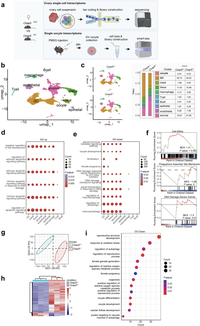
Methods: Bulk RNA sequencing of granulosa cells was conducted in the control group and in the patients with DOR to screen for candidate genes, which were further validated by gene burden analysis in a next-generation sequencing cohort of POI and control individuals. Additionally, ovarian reserve was evaluated in heterozygous Ckap5 knockout mice, alongside the ovarian and oocyte single-cell transcriptome analysis. The regulatory mechanism of CKAP5 was studied through in vivo and in vitro experiments.

Findings: CKAP5 was identified as a key hub gene associated with ovarian ageing. Heterozygous Ckap5 knockout mice exhibited a POI-like phenotype, characterized by a reduced primordial follicle pool and accelerated follicular atresia. CKAP5 promotes autophagy via ATG7 and simultaneously supports DNA damage repair through the ATM. Finally, a variant in CKAP5 (NM_0001008938.4, c.630 + 7_630 + 11delCAAAA) was identified in patients with POI, resulting in protein truncation and loss of function.

Interpretation: CKAP5 deficiency induces premature ovarian insufficiency in both humans and mice.

Funding: The National Key R&D Program of China (2017YFC1001100), the National Natural Science Foundation of China (81501248, 81471453 and 81801295), the Health Research Project of Hunan Provincial Health Commission (W20243018), the Science and Technology Innovation Program of Hunan Province (2021RC3031), the National Natural Science Foundation of Hunan Province (2022JJ30066), the Scientific Research Program of Hunan Provincial Health Commission (202205033471 and 21B0058), the Open Research Fund of Hunan Provincial Key Laboratory of Regional Hereditary Birth Defects Prevention and Control (HPKL2023013).

Keywords: Apoptosis; Autophagy; CKAP5; DNA damage repair; Premature ovary insufficiency.


Conflict of interest statement

Declaration of interests The authors declare no conflict of interest.

Figures

Fig. 1
Fig. 1
Identification of CKAP5 as a hub gene for ovarian ageing. a. The flowchart of identification of CKAP5 as a hub gene for ovarian ageing. b. PCA analysis of RNA transcriptomes in the DOR vs. CON cohort (n = 5 for each group). c. Heatmap of DEGs. Red indicates upregulated genes and blue indicates downregulated genes in the DOR group (n = 5 for each group). d. The cluster dendrogram and hierarchical clustering heatmap of module eigengenes. e. The heatmap of correlation between genes in different modules and the clinical traits through WGCNA analysis. f. Venn diagrams of candidate genes obtained through intersecting the hub genes in turquoise module and DEGs. g. The verification of CKAP5 by qRT‒PCR in the DOR vs. CON cohort (n = 5, Student’s t-test, ∗∗∗∗P < 0.0001). DOR, diminished ovarian reserve; CON, control; CKAP5, Cytoskeleton Associated Protein 5; WGCNA, weighted gene co-expression network analysis; DEGs, differentially expressed genes; PCA, principal component analysis; oocyte, the number of follicles over 14 mm on HCG Day.
Fig. 2
Fig. 2
Ckap5 knockout results in subfertility due to POI in mouse. a. Immunohistochemistry of CKAP5 in ovarian sections from 1-month-old wildtype mice. Scale bars denote 50 μm, 100 μm and 400 μm. b. Immunofluorescence analysis for CKAP5 and α-tubulin colocalization in the oocyte maturation from wildtype adult mice. Scale bars denote 20 μm. c. Schematic diagram of Ckap5 knockout. CRISPR/Cas9 technology was used to generate Ckap5+/− mice by deleting exon 3 to exon 6. d. Genotype of Ckap5+/+ and Ckap5+/− mice. Heterozygotes: two bands with 714 bp and 643 bp. Wild-type allele: one band with 643 bp. e. Western blot analysis of CKAP5 in ovary from Ckap5+/+ and Ckap5+/− mice. f. Relative protein levels of CKAP5 in ovary from Ckap5+/+ and Ckap5+/− mice (n = 3, Student’s t-test, ∗∗P < 0.01). Relative amounts of proteins were calculated after normalization to the α-tubulin protein level. g. Immunohistochemistry analysis of CKAP5 expression in the ovary from Ckap5+/+ and Ckap5+/− mice. Scale bars denote 100 μm. h. The ovarian development of Ckap5+/+ and Ckap5+/− female adult mice. i. The ovary/body weight of Ckap5+/+ and Ckap5+/− female adult mice (n = 3, Student’s t-test, ∗∗P < 0.01). j. The numbers of litters per mouse, the number of pups per litter and the numbers of total pups per mouse were recorded for each group in the fertility test (n = 3, Welch’s t-test, litter/mouse: ∗P < 0.05; Mann–Whitney test, pups/litter: ∗∗∗∗P < 0.0001; Student’s t-test, total pups/mouse: ∗∗∗P < 0.001). k. GV oocytes obtained from Ckap5+/− and Ckap5+/+ mice at 4 and 12 weeks old (n = 3, Student’s t-test, 4w: ∗P < 0.05; Welch’s t-test, 12w: ∗P < 0.05). l. Serum FSH, LH and E2 levels of 3-month-old, 6-month-old and 9-month-old Ckap5+/+ and Ckap5+/− mice (n = 4, 3 and 6 months: Student’s t-test; 9 months: Mann–Whitney test; ∗P < 0.05, ∗∗P < 0.01). m. HE staining and comparison of the numbers of follicles of mice at 1-month-old, 2-month-old, 4-month-old and 6-month-old (n = 3, Student’s t-test, ∗P < 0.05, ∗∗P < 0.01, ∗∗∗P < 0.001, ∗∗∗∗P < 0.0001). Scale bars denote 400 μm. GV, germinal vesicle stages; GVBD, germinal vesicle breakdown stages; MI, metaphase I stages; MII, metaphase II stages; PrF, primordial follicle; PF, primary follicle; SF, secondary follicles; AF, antral follicle; AtF, atresia follicle; FSH, Follicle-stimulating hormone; E2, Oestradiol; LH, Luteinizing hormone.
Fig. 3
Fig. 3
ScRNA-seq analysis reveals the decreased autophagy and DNA damage repair along with increased apoptosis in ovaries of Ckap5+/− mice. a. The flowchart of ovary scRNA-seq and single-oocyte transcriptomic in Ckap5+/− and Ckap5+/+ mice. b. UMAP plot of combined ovarian cells. Clustering analysis revealed 10 distinct ovarian cell populations. c. UMAP plot of ovarian cells by genotype. Numbers and percentages of cell type identity by genotype. d. GO enrichment analysis of genes of up-regulated DEGs in different cell types. e. GO enrichment analysis of genes of down-regulated DEGs in different cell types. f. GSEA enrichment analysis of the oocyte for the pathways related to autophagy, DNA damage repair, and cell killing. g. PCA analysis of oocyte RNA transcriptomes form Ckap5+/− and Ckap5+/+ mice. h. Heatmap of DEGs. Red indicates upregulated genes and blue indicates downregulated genes in the Ckap5+/− group. i. GO enrichment analysis of down-regulated DEGs. GO, gene ontology, GSEA, Gene Set Enrichment Analysis; NES, normalized enrichment score; DEGs, differentially expressed genes.
Fig. 4
Fig. 4
CKAP5 promotes DSBs repair and autophagosome formation to prevent aberrant DNA damage and maintain ovarian autophagy flux. a. Protein levels of CKAP5, ATM, p-ATM, CHK2, p-CHK2, P53, p-P53, SMC3, p-SMC3, SMC1A, p-SMC1A and γ-H2AX in HEK293T cells transfected with siCKAP5 and siControl as measured by Western blot. GAPDH was used as the loading control. b. Protein levels of CKAP5, ATM, p-ATM, CHK2, p-CHK2, P53, p-P53, SMC3, p-SMC3, SMC1A, p-SMC1A and γ-H2AX in HEK293T cells transfected with siCKAP5 and siControl or treated with ETO as measured by Western blot. α-Tubulin was used as the loading control. c. Protein levels of ATM, p-ATM, CHK2, p-CHK2, P53, p-P53, SMC3, p-SMC3, SMC1A, p-SMC1A and γ-H2AX in ovary from Ckap5+/− and Ckap5+/+ mice as measured by Western blot. GAPDH was used as the loading control. d. Immunohistochemistry analysis for ATM, p-ATM and γ-H2AX in the ovary of Ckap5+/+ and Ckap5+/− adult mice. Scale bars denote 100 μm. e. Immunofluorescence analysis for CKAP5 and ATM colocalization in the oocyte maturation from Ckap5+/− and Ckap5+/+ mice. Scale bars denote 20 μm. f. Protein levels of CKAP5, ATG7, ATG5, ATG3, P62 and LC3 in HEK293T cells transfected with siCKAP5 and siControl as measured by Western blot. GAPDH was used as the loading control. g. Immunofluorescence analysis for LC3 expression were decreased in HEK293T cells transfected with siCKAP5. Scale bars denote 200 μm. h. Protein levels of CKAP5, ATG7, ATG5, ATG3, P62 and LC3 in HEK293T cells transfected with siCKAP5 and siControl or pre-treated with EBSS and/or CQ as measured by Western blot. α-Tubulin was used as the loading control. i. Protein levels of CKAP5, ATG7, ATG5, ATG3, P62 and LC3 in HEK293T cells transfected with siCKAP5 and siControl or pre-treated with CQ for 0–4 h as measured by Western blot. α-Tubulin was used as the loading control. j. Protein levels of ATG7, ATG5, ATG3, LC3 and P62 in ovary from Ckap5+/− and Ckap5+/+ mice as measured by Western blot. α-tubulin was used as the loading control. k. Immunohistochemistry analysis for ATG7 and LC3 in the ovary of Ckap5+/+ and Ckap5+/− adult mice. Scale bars denote 100 μm. l. Immunofluorescence analysis for CKAP5 and ATG7 colocalization in the oocyte maturation from Ckap5+/− and Ckap5+/+ mice. Scale bars denote 20 μm. DSBs, double strand breaks; ETO, etoposide; MI, metaphase I stages; MII, metaphase II stages; EBSS, Earle’s Balanced Salt Solution; CQ, Chloroquine.
Fig. 5
Fig. 5
CKAP5 variant causes clinical POI. a. The pedigrees of the families. Squares and circles denote male and female members, respectively. Solid symbols indicate the affected members, and open symbols denote unaffected members. Slashes represent deceased members, and the equal sign indicates infertility. Members indicated by arrows were probands. Members indicated by asterisk were selected for whole-exome sequencing. Sanger sequencing chromatograms of CKAP5 are shown on the bottom side. The red arrows indicate the corresponding variant. b. Schematic map of the variant positions in CKAP5 at the genomic level. c. Mini-gene assay of wild-type and mutated CKAP5 plasmids transfected into HEK293T cells. Sanger sequencing of the band form mini-gene assay. d. Schematic map of the variant positions in CKAP5 at the protein level. e. Protein structure of wild-type and mutated CKAP5 predicted by Alphafold3. f. Wild-type and mutated CKAP5 plasmids were transfected into HEK293T cells. After 72 h of transfection, whole-cell lysates were prepared and separated by SDS-PAGE. CKAP5 was detected by His antibody and GAPDH served as a loading control. Arrows indicate the targeted proteins. g. Protein levels of ATG7, ATG5, ATG3, LC3, P62, ATM, p-ATM, CHK2, p-CHK2, P53, p-P53, SMC3, p-SMC3, SMC1A, p-SMC1A and γ-H2AX in HEK293T cells transfected with wildtype and mutated CKAP5 plasmids as measured by Western blot. α-Tubulin was used as the loading control. WT, wild type; MT, mutant type; con, control.

Similar articles

Cited by

References

    1. Webber L., Davies M., Anderson R., et al. European Society for Human Reproduction and Embryology (ESHRE) Guideline Group on POI ESHRE Guideline: management of women with premature ovarian insufficiency. Hum Reprod. 2016;31(5):926–937. - PubMed
    1. Golezar S., Ramezani Tehrani F., Khazaei S., Ebadi A., Keshavarz Z. The global prevalence of primary ovarian insufficiency and early menopause: a meta-analysis. Climacteric. 2019;22(4):403–411. - PubMed
    1. Ishizuka B. Current understanding of the etiology, symptomatology, and treatment options in premature ovarian insufficiency (POI) Front Endocrinol (Lausanne) 2021;12 - PMC - PubMed
    1. Jiao X., Ke H., Qin Y., Chen Z.J. Molecular genetics of premature ovarian insufficiency. Trends Endocrinol Metab. 2018;29(11):795–807. - PubMed
    1. Qin Y., Jiao X., Simpson J.L., Chen Z.J. Genetics of primary ovarian insufficiency: new developments and opportunities. Hum Reprod Update. 2015;21(6):787–808. - PMC - PubMed