Review
. 2025 Apr;232(4S):S105-S123.
doi: 10.1016/j.ajog.2024.08.043.

Endometriosis and adenomyosis unveiled through single-cell glasses

Affiliations
Review

Endometriosis and adenomyosis unveiled through single-cell glasses

Linda C Giudice et al. Am J Obstet Gynecol. 2025 Apr.

Abstract

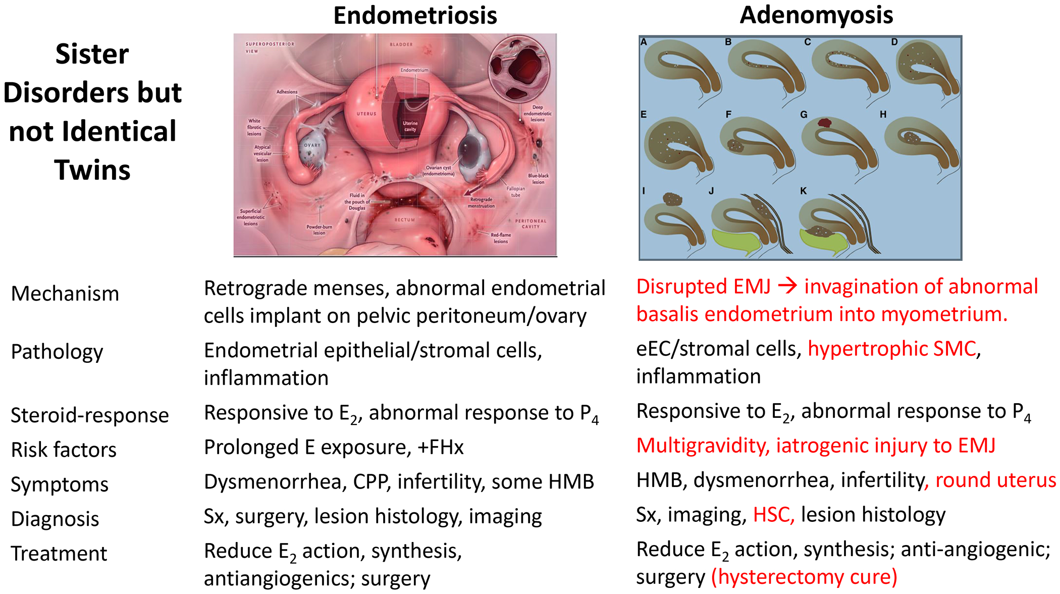
Single-cell technologies are expanding our understanding of endometriosis and adenomyosis, which are sister disorders of the uterine endometrium that contain similar complements of lesion cell types but in different locations-outside and inside the uterus, respectively. Both diseases cause significant morbidity and impaired quality of life among those affected, and current therapies mitigate most of the symptoms although with highly variable efficacy, duration of effect, and frequent intolerable side effects. Thus, there is a pressing need for transformative approaches and to develop individualized therapies for the variety of presentations of endometriosis and adenomyosis symptoms and the heterogeneity of lesion types, both histologically and architecturally. Single-cell technologies are transforming the understanding of human physiology and pathophysiology in the reproductive system and beyond. This manuscript reviews the clinical characteristics of endometriosis and adenomyosis and the recent studies focused on eutopic endometrium and ectopic lesions at single-cell resolution, the myriad of cell types and subtypes, cell-cell communications, signaling pathways, applications for novel drug discovery and therapeutic approaches, and challenges and opportunities that accompany this type of research. Key take-home messages from the studies reviewed herein include the following: We conclude the review with an eye to the future-what Alice might see beyond the single-cell looking glass that connects endometrium and endometrial disorders with the trillions of cells of other tissues and organs in health and disease throughout the human body and the opportunities for novel diagnostic modalities and drug discovery for endometriosis, adenomyosis, and related uterine and inflammatory conditions.

Keywords: adenomyosis; biomarkers; cell types; drug discovery; endometriosis; inflammation; precision medicine; sequencing; signaling pathways; single cell.


Figures

Figure 1.
Figure 1.
Comparison of endometriosis and adenomyosis pathogenesis, risk factors, diagnostic approaches, pathophysiology, symptoms, and therapies. Adapted from Bulun et al, 2023, with permission.
Figure 2.
Figure 2.. Absence of key genes that open the window of implantation in eutopic endometrial epithelial cluster in infertile patients with Stage I/II endometriosis.
Panel A. t-SNE projection of endometrial cells colored based on their sample origin and cycle phase. Panel B. Expression patterns of PAEP and CXCL14 projected on t-distributed stochastic neighbour embedding (t-SNE) plot. From Huang et al, 2023, with permission.
Figure 3.
Figure 3.. Distinct Cell Compositions and Cell Enrichment in Menstrual Endometrium from patients with Endometriosis versus Controls and Model of Disease.
Panels A and B show low abundance of uNK1 and uNK2 cells and high abundance of B cells in patients with endometriosis. Panel C shows proposed pathogenesis and pathophysiology implied by the data. From Shih et al, 2022, with permission.
Figure 4.
Figure 4.. Endometriosis lesion scRNAseq analysis.
Panel A. Histologic (H&E staining) and architectural heterogeneity of endometriosis lesions. Panel B. Correlations Based on Cluster Frequency Across All Specimens Profiled by scRNAseq. Panel C. Epithelial differential gene expression and signaling pathways in endometriomas and superficial and deep disease (P < 0.05 and log2 FC > 1). From Fonseca et al, 2023, with permission.
Figure 5.
Figure 5.. Molecular signatures of epithelial and stromal cells in superficial peritoneal and deep endometriosis differ.
Endometrial-type epithelial cells (Panel A) and fibroblasts (Panel B) exhibit DGE associated with deep or superficial status. From Fonseca et al, 2023, with permission.
Figure 6.
Figure 6.. Endometrium, endometriosis, and endometriomas share somatic mutations with transcriptional consequences.
Panel A. Mutations detected in each lesion. Panel B. Differential gene expression by mutation state. Panel C. Epithelial expression of lymph-angiogenesis markers by ARID1A staining status. From Fonseca et al, 2023, with permission.
Figure 7.
Figure 7.. Deconvolution Analysis Reveals Endometrial-type Epithelium Cell-type Signatures in Endometriosis-associated Ovarian Cancers.
Multi-subject single-cell deconvolution revealed strong enrichment of signatures for ciliated endometrial-type epithelial cells compared with the other keratin-positive clusters. HGSOC, an epithelial ovarian tumor type thought not to originate from endometriosis epithelial cells showed no pattern for preferential enrichment of any epithelial cluster. From Fonseca et al, 2023, with permission.
Figure 8.
Figure 8.. Immune cell frequencies in endometriomas and communication networks of immune cell types with endometrioma fibroblasts.
The network supports interactions between immune and fibroblast cells in ovarian endometrioma pathogenesis and pathophysiology. From Ma et al, 2021, with permission.
Figure 9.
Figure 9.. The endometrial-myometrial junction (EMJ).
Panel A. Histology of human uterine endometrium and myometrium compartments (H&E staining). Panel B: Transvaginal ultrasound midsagittal image of uterine compartments. Panel C. Schematic of uterine compartments. Adapted from Naftlin and Jurkovic, 2009, with permission.
Figure 10.
Figure 10.. Fibroblasts and epithelial cell subtypes differ in endometrium and adenomyosis.
Panel A. UMAP of cell types and fibroblast trajectories in endometrium. B. UMAP of cell types and fibroblast trajectories in adenomyosis. Panel C. Progesterone resistance of epithelial subtypes in adenomyosis. Panel D. WNT signaling pathway network. From Yildiz et al, 2023, with permission.

Similar articles

References

    1. Zondervan KT, Becker CM, Koga K, Missmer SA, Taylor RN, Viganò P. Endometriosis. Nat Rev Dis Primers. 2018;4(1):9. doi: 10.1038/s41572-018-0008-5 - DOI - PubMed
    1. Zondervan KT, Becker CM, Missmer SA. Endometriosis. N Engl J Med. 2020;382(13):1244–1256. doi: 10.1056/NEJMra1810764 - DOI - PubMed
    1. Bulun SE, Yilmaz BD, Sison C, et al. Endometriosis. Endocr Rev. 2019;40(4):1048–1079. doi: 10.1210/er.2018-00242 - DOI - PMC - PubMed
    1. Giudice LC, Oskotsky TT, Falako S, Opoku-Anane J, Sirota M. Endometriosis in the era of precision medicine and impact on sexual and reproductive health across the lifespan and in diverse populations. FASEB Journal. 2023;37(9). doi: 10.1096/fj.202300907 - DOI - PMC - PubMed
    1. Zhai J, Vannuccini S, Petraglia F, Giudice LC. Adenomyosis: Mechanisms and Pathogenesis. Semin Reprod Med. 2020;38(2-03):129–143. doi: 10.1055/s-0040-1716687 - DOI - PMC - PubMed