. 2025 Apr 19;22(1):108.
doi: 10.1186/s12985-025-02729-3.

The first trimester human placenta responds to Zika virus infection inducing an interferon (IFN) and antiviral interferon stimulated gene (ISG) response

Affiliations

The first trimester human placenta responds to Zika virus infection inducing an interferon (IFN) and antiviral interferon stimulated gene (ISG) response

Kylie H Van der Hoek et al. Virol J. .

Abstract

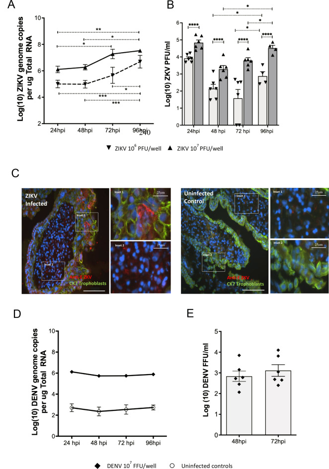
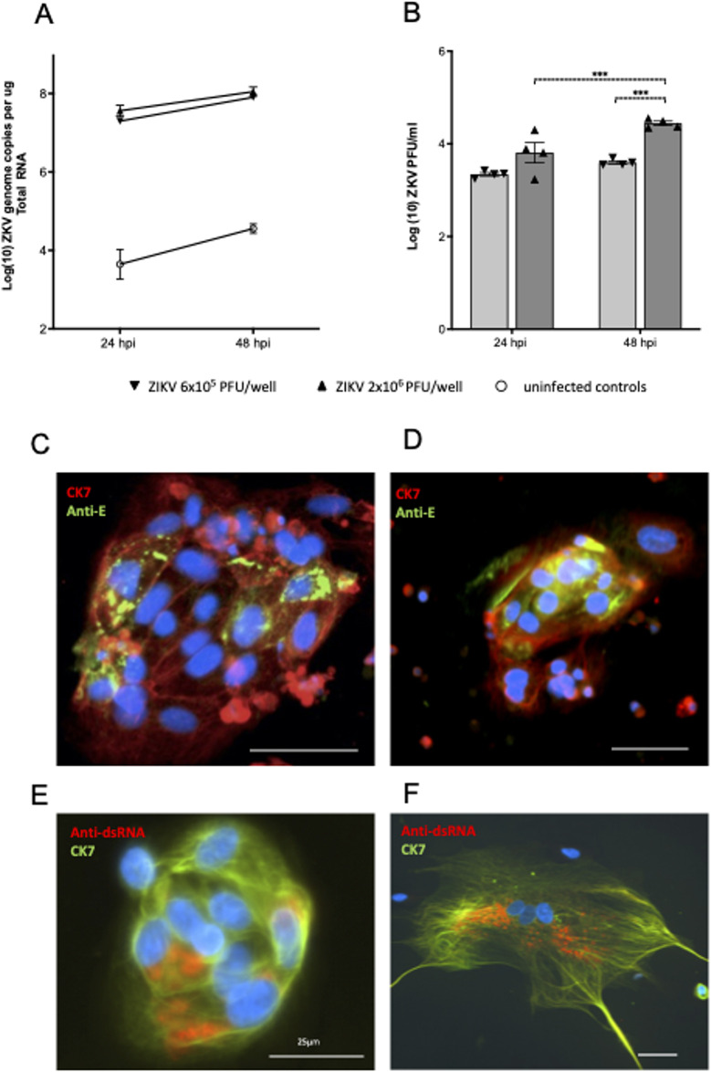
Background: Zika virus (ZIKV) is a positive-strand RNA virus of the Flaviviridae family. Maternal ZIKV infection during pregnancy can spread to the placenta and fetus causing severe neurological defects and infants born with microcephaly. Here, we investigated ZIKV infection and the cellular innate antiviral immune response in first trimester human placental explant cultures and isolated primary villus cytotrophoblasts (CTBs).

Methods: Placentas were obtained with informed consent from women undergoing elective pregnancy termination and either cultured as placental explants or used to isolate primary CTBs. Explants and CTBs were both infected with ZIKV (PRVABC59), and samples evaluated for infection by qRT-PCR, viral plaque and ELISA assays, and immunohistochemical or immunocytochemical staining.

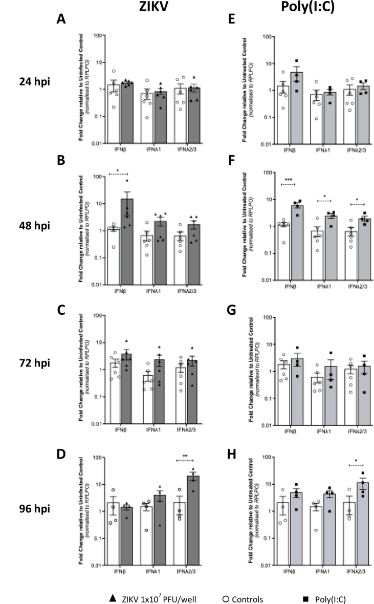
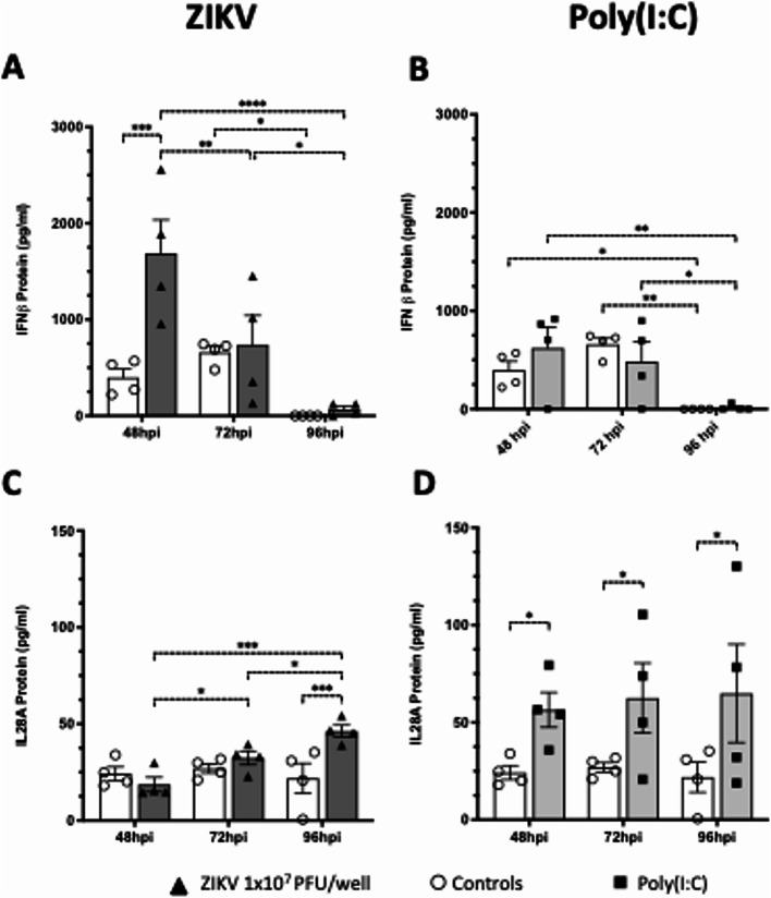
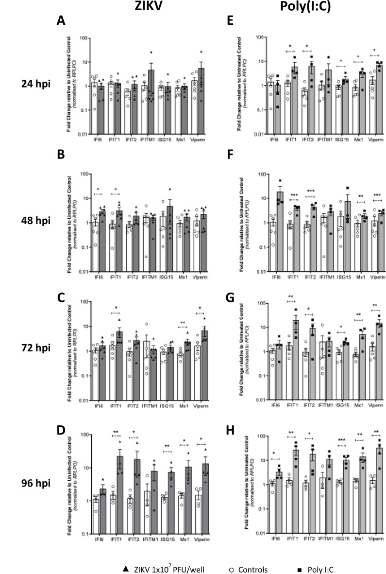
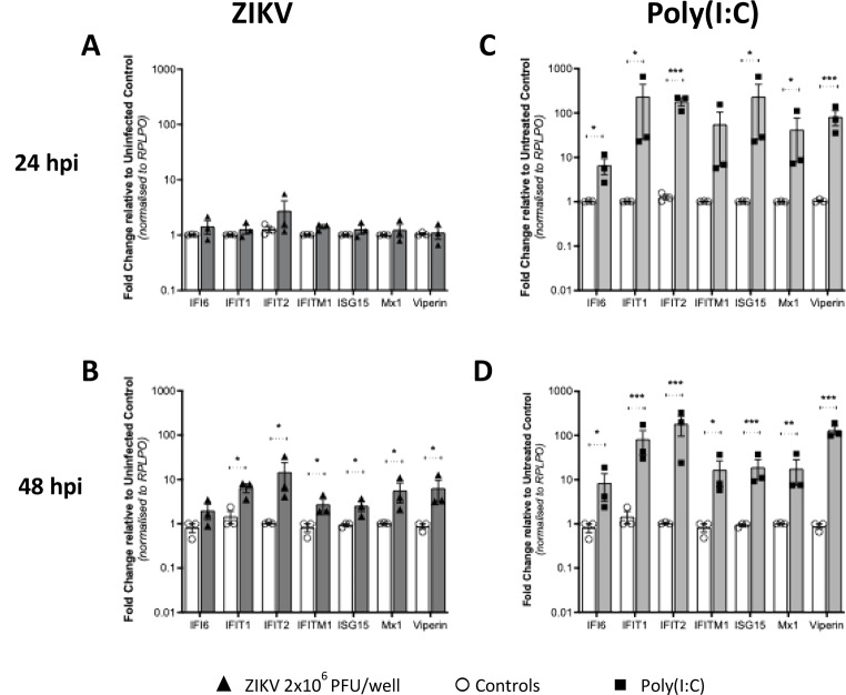
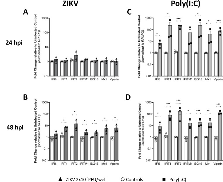
Results: We demonstrate robust infection and production of ZIKV in placental explant and CTB cultures. Both displayed delayed upregulation of interferons (IFN), most notably IFNβ and IFNλ2/3, and a panel of interferon stimulated genes (ISG) (IFI6, IFIT1, IFIT2, IFITM1, ISG15, MX1, RSAD). Stimulation of explants and CTBs with the dsRNA mimic poly(I: C), caused immediate IFN and ISG upregulation, demonstrating the first trimester placenta is innate immune competent. This suggests that either ZIKV blocks the early innate response, or the placental response is inherently hindered.

Conclusion: Together these data show that first trimester placenta is susceptible to ZIKV infection which induces a delayed type III IFN antiviral response. This delay likely creates an environment favourable to ZIKV replication and dissemination across the early gestation placenta to fetal tissue, causing pathologies associated with congenital ZIKV syndrome.

Keywords: First trimester pregnancy; Interferon; Interferon lambda; Interferon stimulated genes; Placenta; Trophoblast; Zika virus.


Conflict of interest statement

Declarations. Ethics approval and consent: All placentas were obtained with written informed consent from women undergoing elective pregnancy terminations at the Pregnancy Advisory Centre (PAC), Woodville, South Australia. Ethics approval was granted by the Queen Elizabeth Hospital Human Research Ethics Committee (HREC/16/TQEH/33). Competing interests: The authors declare no competing interests.

Figures

Fig. 1
Fig. 1
ZIKV infects and replicates in first trimester placental explants. First trimester placental explant tissue cultures (n = 6 patients in duplicate) were infected with ZIKV at 106 or 107 PFU/well and (A) ZIKV RNA genomes quantified by qRT-PCR. Data were log transformed and analysed by Ordinary Two Way ANOVA (* P < 0.05, ** P < 0.01. *** P < 0.005) or (B) infectious ZIKV particles in culture supernatant from infected explant tissues detected by plaque assay. Some explants that were infected at 106 PFU/well did not produce infectious plaques at 72hpi. The 24hpi data represents the viral inoculum, as virus was left on explants for 24 h. This data was not included in subsequent statistical analysis. Data were log transformed and analysed by Ordinary Two-Way ANOVA. (* P < 0.05, **** P < 0.0001). (C) Frozen sections from ZIKV infected explant tissues (96hpi) were stained for ZIKV-Envelope antigen (red), cytokeratin (green) and nuclei (DAPI). Bars represent 100 μm unless otherwise indicated. Images are representative of multiple samples analysed (D). First trimester placental explant tissue cultures (6 patients in duplicate) were infected with DENV and at timepoints indicated DENV RNA genomes quantified by qRT-PCR including in uninfected controls. (E) infectious DENV particles in the culture supernatant from infected explant tissues detected by focus forming assay. All data are means± SE
Fig. 2
Fig. 2
Zika virus infects and replicates in isolated first trimester placental trophoblasts. (A) Trophoblasts from first trimester placental samples were infected with ZIKV then at 24hpi and 48hpi ZIKV genome RNA was quantified using qRT-PCR, including in uninfected control cells. Data are means ± SE, n = 3 patient samples. (B) Infectious ZIKV in CTB culture supernatant was quantified by plaque-assay. Data are means ± SE, n = 2 patient samples in duplicate. Data were log transformed and analysed by Ordinary Two Way ANOVA (*** P < 0.005). (C-D) Isolated trophoblasts were fixed and stained for the CTB marker, cytokeratin-7 (red) and ZIKV-E antigen (green). (E, F) Detection of ZIKV genome dsRNA replication intermediates used anti-dsRNA Ab (red), cytokeratin 7 (green) and nuclei (DAPI). Bars represent 50 μm unless otherwise indicated
Fig. 3
Fig. 3
ZIKV infection induces a Type III IFN response in first trimester placental explant tissue. Explant tissue was (A, B,C, D) infected with 107/ml ZIKV or (E, F,G, H) stimulated with 1 µg/ml poly(I: C)(A, B,C are n = 6 patients) (D, E,F, G,H are n = 4 patients). RNA was collected 24, 48, 72 and 96hpi followed by qRT-PCR analysis for the detection of IFNβ, IFNλ1 and IFNλ2/3. Data are normalised to the housekeeping gene RPLP0 and expressed as a fold-change relative to mock-infected control at each timepoint. (data are means ± SE) * P < 0.05, ** P < 0.01 ***< P0.005. Statistical analysis was performed on log transformed data at each time point using multiple unpaired T test for each gene
Fig. 4
Fig. 4
ZIKV infection induces an IFN protein in response in first trimester placental explant tissue. Explant tissue was infected with 107/ml ZIKV (A, C) or stimulated with 1 µg/ml poly(I: C) (B, D) (n = 4 patients, data are means ± SE. *P < 0.05, **P < 0.01, ***P < 0.005, ****P < 0.0001). Conditioned culture media was collected from cultured explants at 48, 72 and 96hpi and then analysed by ELISA assay for hIFNβ and hIL-28 A (I FNλ2). Statistical analysis was performed using a 2-way ANOVA
Fig. 5
Fig. 5
ISG responses in first trimester placental explant tissue. Explant tissue was (A, B,C, D) infected with 107/ml ZIKV or (E, F,G, H) stimulated with 1 µg/ml poly(I: C)(A, B,C are n = 6 patients, D, E,F, G,H are n = 4 patients. RNA was collected 24, 48, 72 and 96hpi followed by qRT-PCR analysis for the detection of antiviral ISGs. Data are normalised to the housekeeping gene RPLP0 and expressed as a fold-change relative to mock-infected control at each timepoint (data are means ± SE) * P < 0.05, ** P < 0.01 ***< P0.005. Statistical analysis was performed on log transformed data at each time point using multiple unpaired T test for each gene
Fig. 6
Fig. 6
Interferon mRNA expression in isolated first trimester placental trophoblasts. Trophoblasts isolated from first trimester placental samples were (A and B) infected with ZIKV MOI-5 or (C and D) stimulated with 1 µg/ml poly(I: C) and RNA collected at 24 h and 48 h (n = 3 patient samples in duplicate). qRT-PCR was then used to detect IFNβ, IFNλ1 and IFNλ2/3 mRNA. Data are normalised to the housekeeping gene RPLP0 and expressed as a fold-change relative to the mock-infected control at each timepoint (data are means ± SE). Statistical analysis was performed on log transformed data at each time point using multiple unpaired T test for each gene
Fig. 7
Fig. 7
ISGs mRNA expression in isolated first trimester placental trophoblasts. Trophoblasts isolated from first trimester placental samples were (A and B) infected with MOI-5 ZIKV or (C and D) stimulated with 1 µg/ml poly(I: C) and RNA collected at 24 h and 48 h (n = 3 patients). qRT-PCR was then used to detect mRNA for antiviral ISGs. Data are normalised to the housekeeping gene RPLP0 and expressed as a fold-change relative to the mock-infected control at each timepoint (data are means ± SE). Statistical analysis was performed on log transformed data at each time point using multiple unpaired T test for each gene

Similar articles

References

    1. Brasil P, Pereira JP Jr., Moreira ME, Ribeiro Nogueira RM, Damasceno L, Wakimoto M, et al. Zika virus infection in pregnant women in Rio de Janeiro. N Engl J Med. 2016;375(24):2321–34. - PMC - PubMed
    1. Antoniou E, Orovou E, Sarella A, Iliadou M, Rigas N, Palaska E et al. Zika virus and the risk of developing microcephaly in infants: A systematic review. Int J Environ Res Public Health. 2020;17(11). - PMC - PubMed
    1. Martins MM, Alves da Cunha AJL, Robaina JR, Raymundo CE, Barbosa AP, Medronho RA. Fetal, neonatal, and infant outcomes associated with maternal Zika virus infection during pregnancy: A systematic review and meta-analysis. PLoS ONE. 2021;16(2):e0246643. - PMC - PubMed
    1. Mulkey SB, Arroyave-Wessel M, Peyton C, Bulas DI, Fourzali Y, Jiang J et al. Neurodevelopmental abnormalities in children with in utero Zika virus exposure without congenital Zika syndrome. JAMA Pediatr. 2020. - PMC - PubMed
    1. Gazeta RE, Bertozzi A, Dezena R, Silva ACB, Fajardo TCG, Catalan DT et al. Three-Year clinical Follow-Up of children intrauterine exposed to Zika virus. Viruses. 2021;13(3). - PMC - PubMed

Publication types