. 2025 Apr 19;23(1):458.
doi: 10.1186/s12967-025-06437-y.

Peptides based on the interface of hnRNPA2B1-transthyretin complex repress retinal angiogenesis in diabetic retinopathy

Affiliations

Peptides based on the interface of hnRNPA2B1-transthyretin complex repress retinal angiogenesis in diabetic retinopathy

Yixiu Chen et al. J Transl Med. .

Abstract

Background: Heterogeneous nuclear ribonucleoprotein A2B1 (hnRNPA2B1) plays a vital role in angiogenesis, when its nucleic acid-binding domain is occupied by transthyretin (TTR), the neovascularization of human retinal microvascular endothelial cells (hRECs) is repressed under hyperglycemic conditions.

Methods: HnRNPA2B1-targeting peptides (THIPs) were designed based on the core fragments at the TTR-hnRNPA2B1 interface. Biacore, Langmuir equilibrium adsorption, and co-immunoprecipitation (co-IP) assays were performed to determine the association between the THIPs and hnRNPA2B1. Proliferation and DNA synthesis in hRECs were detected using CCK-8 and EdU assays. Transwell, wound healing, and tube formation assays were used to evaluate migratory and the angiogenic capacity of hRECs. Related RNA and protein expression levels were tested by quantitative PCR and western blot assays, respectively. Streptozotocin (STZ)-induced diabetic retinopathy (DR) model rats were intravitreally injected with 5 μL of AAV9 virus (1 × 10¹² vg/mL) every 8 weeks, with sterile saline used as control. After 16 weeks, the retinas were extracted and subjected to Evans blue leakage and retinal trypsin digestion assays. Retinal paraffin sections were prepared and stained with hematoxylin and eosin (H&E) or subjected to immunohistochemical or immunofluorescence assays.

Results: Biacore, Langmuir equilibrium adsorption, and co-IP analyses demonstrated that the four designed THIPs specifically recognized hnRNPA2B1. CCK-8 and EdU labeling assays showed that the THIPs inhibited proliferation and DNA synthesis in hRECs under hyperglycemia. Transwell, wound healing and tube formation assays demonstrated that the THIPs inhibited the migratory and angiogenic capacity of hRECs. Quantitative PCR and western blot assays suggested that the THIPs exerted their effects via the STAT4/miR-223-3p/FBXW7 and the downstream Notch1/Akt/mTOR axes. In vivo studies using DR model rat revealed that the intravitreal administration of THIP-4 significantly mitigated retinal leakage, capillary decellularization, pericyte loss, fibrosis, and gliosis during DR progression.

Conclusion: Our findings demonstrated that under hyperglycemia, THIP-4 suppressed DR progression via the STAT4/miR-223-3p/FBXW7 and Notch1/Akt/mTOR axes both in vitro and in vivo. These results indicated that THIP-4 has strong potential for clinical application in DR and other angiogenesis associated diseases.

Keywords: DR rat model; Diabetic retinopathy; HnRNPA2B1; Retinal angiogenesis; THIPs.


Conflict of interest statement

Declarations. Ethics approval and consent to participate: The study was approved by the Ethics Committee of the Affiliated Wuxi People’s Hospital of Nanjing Medical University (2022-033, 18-Oct-2022) in China. Consent for publication: Not applicable. Competing interests: The authors declare no competing interests.

Figures

Fig. 1
Fig. 1
The design of TTR-hnRNPA2B1 interface peptides (THIPs). A The TTR homotetramer (marked in green)/hnRNPA2B1 (marked in purple) complex; the interface is marked in yellow. Two amino acid substitutions—I68R and D99R—are marked in red. B The sequences of the THIPs and the scramble peptide. C Following the lysis of human retinal microvascular epithelial cells (hRECs), anti-hnRNPA2B1 and anti-His-tag antibodies were used to pull down the hnRNPA2B1-THIPs complexes, and these were then subjected to western blot or dot blot analysis. D After 2 h of incubation with 1 μM THIPs, TTR, or scrambled peptide, the localization of these molecules was detected using anti-hnRNPA2B1, anti-human TTR, anti-His-tag, Alexa Fluor 488-labeled anti-IgG, and Alexa Fluor 594-labeled anti-IgG antibodies; nuclei were counterstained with Hoechst 33342. Scale bar: 25 μm
Fig. 2
Fig. 2
The THIPs inhibited DNA synthesis and proliferation of hRECs. (A, B) EdU assays (1, vs. HG; 2, vs. HG + NC; 3, vs. HG + THIP-4; n = 5, *P < 0.05, **P < 0.01, ***P < 0.001); nuclei were counterstained with Hoechst 33342, scale bar: 100 μm. (C) CCK-8 assays after 5 days of culture (1, vs. HG; 2, vs. HG + NC; 3, vs. HG + THIP-4; n = 5, *P < 0.05, **P < 0.01, ***P < 0.001). HG high glucose, NC negative control
Fig. 3
Fig. 3
The THIPs inhibited migration, wound healing, and tube formation of hRECs. A Transwell assay (32 h; scale bar: 500 μm). B, C Wound healing assay (24 h; 1, vs. HG; 2, vs. HG + NC; 3, vs. HG + THIP-4; n = 5, *P < 0.05, **P < 0.01, ***P < 0.001); scale bar: 200 μm. D, E Tube formation assay (4 h; 1, vs. HG; 2, vs. HG + NC; 3, vs. HG + THIP-4; n = 5, *P < 0.05, **P < 0.01, ***P < 0.001); scale bar: 200 μm. HG high glucose, NC negative control
Fig. 4
Fig. 4
The THIPs regulated the STAT4/hsa-miR-223-3p/FBXW7 and Notch1/Akt/mTOR pathways in hRECs. AC The expression levels of STAT4 mRNA, hsa-miR-223-3p, and FBXW7 mRNA (1, vs. HG; 2, vs. HG + NC; 3, vs. HG + THIP-4; n = 5, *P < 0.05, **P < 0.01, ***P < 0.001). D Western blot assay. EI The relative protein levels of STAT4 and p-STAT4 (1, vs. HG STAT4; 2, vs. HG p-STAT4; 3, vs. HG + NC STAT4; 4, vs. HG + NC p-STAT4; n = 3, *P < 0.05, **P < 0.01, ***P < 0.001), FBXW7, Notch1, p-AKT, and p-mTOR (1, vs. HG; 2, vs. HG + NC; n = 3, *P < 0.05, **P < 0.01, ***P < 0.001). HG high glucose, NC negative control
Fig. 5
Fig. 5
THIP-4 mitigated the progression of DR in a rat model. A Evans blue (EB) stained retinas were visualized with the EVOS FL Auto Imaging System using a reverse-phase setting, where vascular and leakage appeared in dark shadow (scale bar: 100 μm). B Retinal vascular leakage (n = 5 visual fields in three retinas; 1, vs. normal control; 2, vs. STZ + NC; *P < 0.05, **P < 0.01, ***P < 0.001). C Retinal vascular leakage quantified by serum albumin western blotting (n = 3; 1, vs. normal control; 2, vs. STZ + NC; *P < 0.05, **P < 0.01, ***P < 0.001). D Retinas were digested with trypsin, following which the retinal vasculature was subjected to periodic acid-Schiff staining (scale bar: 25 μm; red arrows: acellular capillaries; green arrows: retained pericytes; blue arrows: lost pericytes). E Acellular capillary number and (F) human retinal microvascular epithelial cell (hREC)/pericyte ratio (n = 5, 1, vs. normal control; 2, vs. STZ + NC; *P < 0.05, **P < 0.01, ***P < 0.001). STZ streptozotocin, NC negative control
Fig. 6
Fig. 6
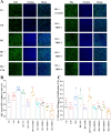
Immunohistochemical assays of the rat retina. A Retinal Sects. (5 μm thick) were subjected to hematoxylin and eosin (H&E) staining (scale bar: 200 μm. GCL, ganglion cell layer; IPL, inner plexiform layer; INL, inner nuclear layer; OPL, outer plexiform layer; ONL, outer nuclear layer; RPE, retinal pigment epithelium) or immunohistochemistry targeting fibronectin, PCNA, HIF-1α, or VEGFA (scale bar: 100 μm). BE The relative positivity ratio for fibronectin, PCNA, HIF-1α, and VEGFA (n = 3; 1, vs. normal control; 2, vs. STZ + NC; *P < 0.05, **P < 0.01, ***P < 0.001). STZ streptozotocin, NC negative control
Fig. 7
Fig. 7
Immunofluorescence assays of the rat retina. Immunostaining for (A) GFAP/GS and (B) TTR/His-tagged THIP-4 on retinal paraffin sections (scale bar: 100 μm). GCL, ganglion cell layer; IPL, inner plexiform layer; INL, inner nuclear layer; OPL, outer plexiform layer; ONL, outer nuclear layer; RPE, retinal pigment epithelium. CF The relative positivity ratio for GFAP, GS, TTR, and His-tagged THIP-4 in the retina (n = 3; 1, vs. normal control; 2, vs. STZ + NC; *P < 0.05, **P < 0.01, ***P < 0.001). STZ streptozotocin, NC negative control
Fig. 8
Fig. 8
THIP-4 regulated the STAT4/rno-miR-223-3p/FBXW7 and Notch1/Akt/mTOR pathways in a DR rat model. A The expression level of rno-miR-223-3p in the rat retina (1, vs. normal control; 2, vs. STZ + NC; n = 3, *P < 0.05, **P < 0.01, ***P < 0.001). B Western blot assay of the proteins extracted from retinal tissue. CG The relative protein levels of STAT4 and p-STAT4 (1, vs. normal control STAT4; 2, vs. normal control p-STAT4; 3, vs. STZ + NC STAT4; 4, vs. STZ + NC p-STAT4; n = 3, *P < 0.05, **P < 0.01, ***P < 0.001), FBXW7, Notch1, p-AKT, and p-mTOR (1, vs. normal control; 2, vs. STZ + NC; n = 3, *P < 0.05, **P < 0.01, ***P < 0.001). STZ streptozotocin; NC negative control
Fig. 9
Fig. 9
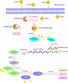
The mechanism underlying the anti-angiogenic function of the TTR-hnRNPA2B1 interface peptides (THIPs)

References

    1. Chen Y, Lundeen EA, Koyama AK, Kompaniyets L, Andes LJ, Benoit SR, Imperatore G, Rolka DB. Prevalence of testing for diabetes among US adults with overweight or obesity, 2016–2019. Prev Chronic Dis. 2023;20:E116. - PMC - PubMed
    1. Kautzky-Willer A, Leutner M, Harreiter J. Sex differences in type 2 diabetes. Diabetologia. 2023;66(6):986–1002. - PMC - PubMed
    1. Krastev E, Abanos S, Kovachev P, Tcharaktchiev D. Diabetes prevalence and duration data extracted from outpatient records representative for the Bulgarian population. Stud Health Technol Inform. 2023;305:230–3. - PubMed
    1. Lundeen EA, Burke-Conte Z, Rein DB, Wittenborn JS, Saaddine J, Lee AY, Flaxman AD. Prevalence of diabetic retinopathy in the US in 2021. JAMA Ophthalmol. 2023;141(8):747–54. - PMC - PubMed
    1. Hou XH, Wang LM, Zhu DL, Guo LX, Weng Ji, Zhang M, Zhou ZG, Zou DJ, Ji QH, Guo XH, et al. Prevalence of diabetic retinopathy and vision-threatening diabetic retinopathy in adults with diabetes in China. Nat Commun. 2023; 14(1). - PMC - PubMed

MeSH terms