. 2025 Apr 19;16(1):3725.
doi: 10.1038/s41467-025-58441-x.

Structural basis for Polθ-helicase DNA binding and microhomology-mediated end-joining

Affiliations

Structural basis for Polθ-helicase DNA binding and microhomology-mediated end-joining

Fumiaki Ito et al. Nat Commun. .

Abstract

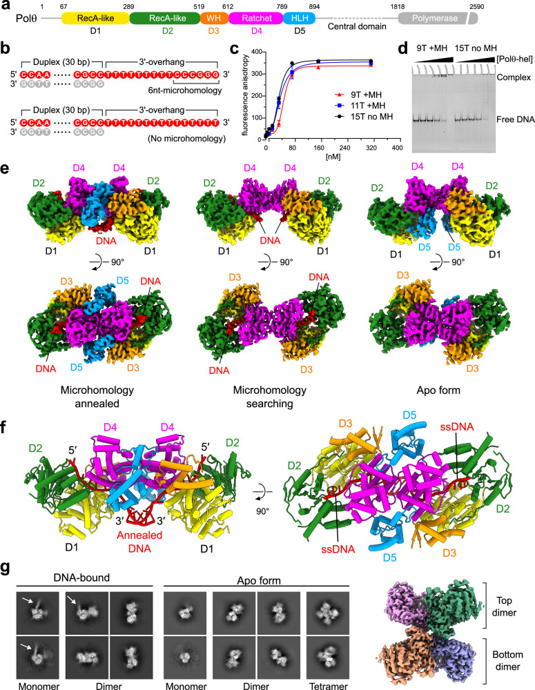
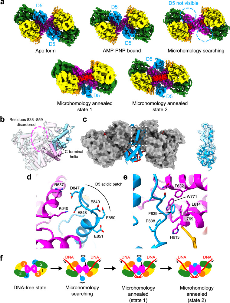
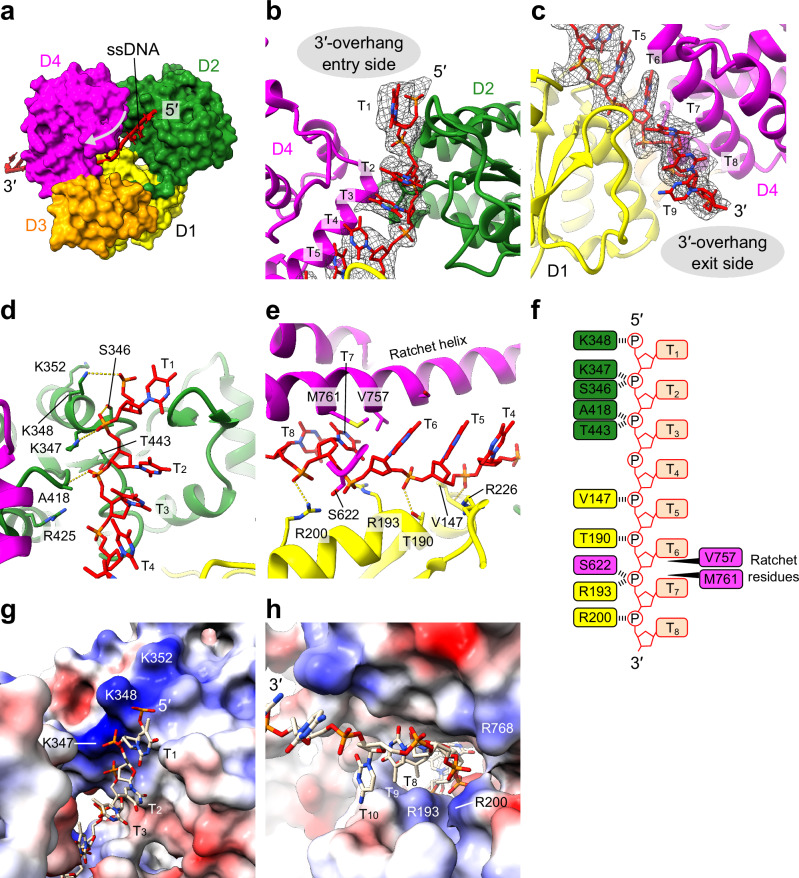
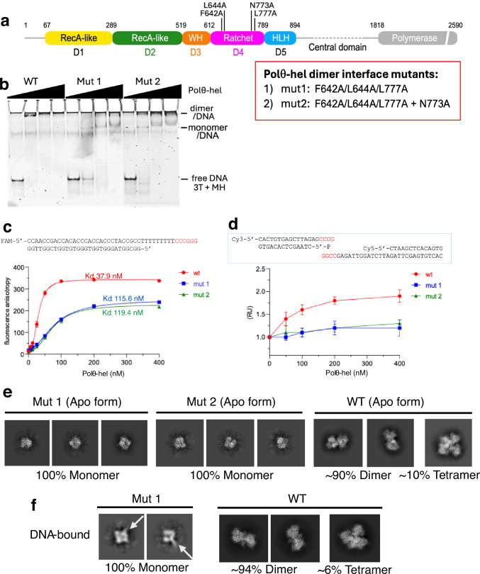
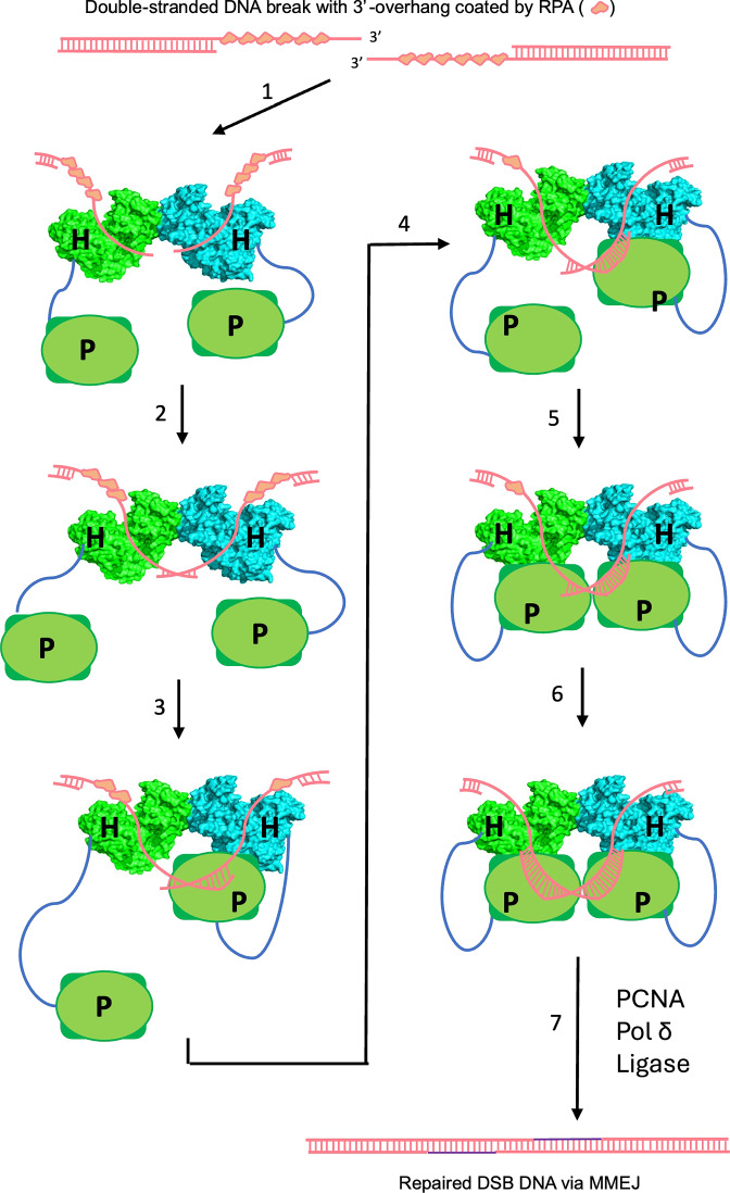
DNA double-strand breaks (DSBs) present a critical threat to genomic integrity, often precipitating genomic instability and oncogenesis. Repair of DSBs predominantly occurs through homologous recombination (HR) and non-homologous end joining (NHEJ). In HR-deficient cells, DNA polymerase theta (Polθ) becomes critical for DSB repair via microhomology-mediated end joining (MMEJ), also termed theta-mediated end joining (TMEJ). Thus, Polθ is synthetically lethal with BRCA1/2 and other HR factors, underscoring its potential as a therapeutic target in HR-deficient cancers. However, the molecular mechanisms governing Polθ-mediated MMEJ remain poorly understood. Here we present a series of cryo-electron microscopy structures of the Polθ helicase domain (Polθ-hel) in complex with DNA containing different 3'-ssDNA overhangs. The structures reveal the sequential conformations adopted by Polθ-hel during the critical phases of DNA binding, microhomology searching, and microhomology annealing. The stepwise conformational changes within the Polθ-hel subdomains and its functional dimeric state are pivotal for aligning the 3'-ssDNA overhangs, facilitating the microhomology search and subsequent annealing necessary for DSB repair via MMEJ. Our findings illustrate the essential molecular switches within Polθ-hel that orchestrate the MMEJ process in DSB repair, laying the groundwork for the development of targeted therapies against the Polθ-hel.


Conflict of interest statement

Competing interests: X.S.C. is a cofounder of Recombination Therapeutics, LLC. R.T.P. is a cofounder and CSO of Recombination Therapeutics, LLC. The remaining authors declare no competing interests.

Figures

Fig. 1
Fig. 1. Architecture of Polθ-hel-DNA complex.
a Schematic representation of the domain organization and construct design of Polθ-hel. WH winged helix, HLH helix–loop–helix. The regions either invisible in any of the cryo-EM maps or excluded from the construct are colored in grey. b DNA substrate sequences used in this study. A 30-bp DNA duplex with a poly(T) 3′-overhang ssDNA was prepared either with 6-nt MH sequence (CCCGGG) or without MH sequence. c Quantitation of DNA binding of Polθ-hel by fluorescence anisotropy. The FAM-labeled DNA with 3′-ssDNA overhang containing 9-nt poly(T) with MH, 11-nt poly(T) with MH, and 15-nt poly(T) without MH were used for the binding assay. Data represent the mean of three technical replicates. n = 3 ± s.d. d EMSA with DNA with 3′-ssDNA overhang containing 9-nt poly(T) with MH and 15-nt poly(T) without MH. The assays were repeated at least three times and all the replicates showed similar results. e Orthogonal views of the cryo-EM maps of Polθ-hel dimer in complex with 3′-ssDNA overhang with 9-nt poly(T) with MH (left), 15-nt poly(T) without MH (middle), and in apo form (right). f Orthogonal views of the atomic model of the Polθ-hel in MH annealed state. g Representative 2D class averages from the data sets of the complex with 15-nt poly(T) DNA without MH 3′-ssDNA overhang (left) and apo form (middle). The cryo-EM map of the apo form tetramer is shown on the right.
Fig. 2
Fig. 2. Structural variability of the mobile domain D5.
a Cryo-EM density maps of Polθ-hel in the apo form, AMP–PNP-bound form, MH search state, and MH annealed states 1 and 2. The subdomain D5 (highlighted in blue) is bound to the helicase ring at the D2–D4 interface in the apo and AMP–PNP-bound forms but becomes invisible in the MH search state. In the MH annealed states, the D5 reemerges at the dimer interface in either one protomer (state 1) or both protomers (state 2). b Superimposition of a protomer from the apo form with the MH annealed state, highlighting the displacement of D5. The C-terminal end of the C-terminal helix is marked with an asterisk. c Positioning of the relocated D5 in the dimer context, depicted in a cylinder model (blue) against the rest of the Polθ-hel dimer shown in the surface representation (grey). An overlay of the cryo-EM density over the atomic model of the relocated D5 is shown on the right. d Structure around the acidic patch of the U-shape structure in D5. Basic residues R637 and K640 of D4 in the other protomer electrostatically engage with the acidic patch. e Structure around F839 of the U-shape structure in D5. The F839 is bound at the hydrophobic cavity formed by L614, F632, L769, and W771 of D4 in the other protomer. f Proposed model of the mobile D5 during the MMEJ.
Fig. 3
Fig. 3. DNA-helicase channel interactions.
a Overview of the 3′-overhang ssDNA (rendered as tube/slabs) threading through the helicase channel (surface model color-coded by subdomains). The direction of DNA translocation is indicated by an arrow. b, c Structure of ssDNA across the helicase channel, showing the atomic model of ssDNA (sticks) near the channel entrance (b) and exit (c), overlaid with the corresponding cryo-EM density (mesh). d Detailed DNA-protein interactions around the channel entrance featuring the 3′-overhang DNA nucleotides T1–T4 (sticks) with surrounding amino acids (ribbons/sticks). e Detailed DNA–protein interactions around the channel exit featuring the 3′-overhang DNA nucleotides T4–T8 (sticks) with surrounding amino acids (ribbons/sticks). f Schematic of the ssDNA strand inside the Polθ-hel channel and interactions with residues from subdomains D1, D2, and D4. g, h Surface electrostatic potential of Polθ-hel. The positively charged patches near channel entrance (g) and exit (h) responsible for ssDNA capture. The surface area is colored according to the calculated electrostatic potential from −10.0 kT/e (red) to +10.0 kT/e (blue).
Fig. 4
Fig. 4. DNA-induced conformational changes in Polθ-hel dimer.
a Superimposition of the Polθ-hel dimer structure in the apo form (cylinder model in light pink) and the complexes with DNA in MH search state (light blue, left) and MH annealed state (lime, right). The bound DNA and the mobile domain D5 have been omitted from the models for clarity. b Superimposition of the Polθ-hel DNA complex in MH annealed state 1 dimer form with the dimer unit of the apo form tetramer. A steric clash between the DNA-bound dimer and the other dimer unit of the apo form tetramer is highlighted. c Superimposition of the apo form (ribbon model in light pink) and the MH annealed state (lime) near the exit of the helicase channel. The movement of the arginine helix in D1 is indicated by arrows. The ratchet helix was used for alignment in the superimposition.
Fig. 5
Fig. 5. Mutant monomeric Polθ-hel is deficient in MMEJ.
a Schematic representation of the domain organization of Polθ and the design of dimer-disrupting Polθ-hel mutants. The positions of the mutated amino acids at the dimer interface and the resulting two mutants (mut 1 and 2) are indicated. b EMSA with WT and mutant Polθ-hel and unlabeled dsDNA with 3′-ssDNA overhangs with 6 nt microhomology (MH) sequence. WT results in a dimer/DNA complex, while mutants 1 and 2 form a monomer/DNA complex. The assays were repeated at least three times and all the replicates showed similar results. c Schematic of DNA used for fluorescence anisotropy (top), which contains a 5′ FAM and a 3′ terminal 6 nt MH sequence (colored in red). Scatter plot shows fluorescence anisotropy for Polθ-hel WT and mutant proteins (bottom). Data represent the mean of three technical replicates. n = 3 ± s.d. d Schematic of DNA used for FRET with Cy3 and Cy5 conjugated to the indicated DNA containing 3′-terminal 4 nt MH. Scatter plot shows relative FRET for Polθ-hel WT and mutant proteins (bottom). Data represent the mean of three technical replicates. n = 3 ± s.d. RU relative units. e Representative 2D class averages from the data sets of the apo proteins of the mutant 1 (left), mutant 2 (middle), and WT (right). f Representative 2D class averages from the data sets of the DNA-bound particles of mutant 1 (left) and WT (right) in the presence of DNA. The bound-DNA is indicated by arrow. The monomer is the only oligomeric form observed in mutant 1, whereas WT forms predominantly dimers, with a smaller fraction of tetramers in the absence of bound DNA.
Fig. 6
Fig. 6. A model of Polθ-hel roles at the initial steps in MMEJ.
The two ends of double-stranded DNA breaks (DSBs) are shown at the top, with RPA binding to the resected 3′-ssDNA overhangs. Polθ forms a dimer via Polθ-hel (each protomer labeled as “H” and colored in green and cyan), with Polθ-pol (labeled as “P”) tethered to the helicase dimer through the flexible Polθ-ctr (labeled as “C”). Step 1: Each protomer of the Polθ-hel dimer binds to one of the two resected 3′-ssDNA overhangs, bringing the opposing 3′-ssDNA ends close to each other at the dimer cleft, allowing for microhomology search and annealing. Polθ-hel ATPase activity can also displace RPA from 3′-ssDNA overhangs. Step 2: Polθ-hel samples the ssDNA sequence microhomology by translocating along the ssDNA and anneals the ssDNA ends at a location with sufficient microhomology. Steps 3, 4: One Polθ-pol protomer could then bind to the short annealed microhomology dsDNA, and extend the primer. Step 5, 6: When the first Polθ-pol protomer extends the primer to a certain length, the second Polθ-pol can bind and extend the opposing primer in the opposite direction to complete the polymerization step. Step 7: In addition to Polθ, Polδ, PCNA, DNA ligase, and other protein factors promote final processing and sealing of the DSB to complete MMEJ repair.

Update of

Similar articles

Cited by

  • Making 3' ends meet.
    Doublié S. Doublié S. Nat Struct Mol Biol. 2025 Jun;32(6):959-960. doi: 10.1038/s41594-025-01578-6. Nat Struct Mol Biol. 2025. PMID: 40404983 No abstract available.

References

    1. Zhao, B., Rothenberg, E., Ramsden, D. A. & Lieber, M. R. The molecular basis and disease relevance of non-homologous DNA end joining. Nat. Rev. Mol. Cell Biol.21, 765–781 (2020). - PMC - PubMed
    1. Jensen, R. B. & Rothenberg, E. Preserving genome integrity in human cells via DNA double-strand break repair. Mol. Biol. Cell31, 859–865 (2020). - PMC - PubMed
    1. Ramsden, D. A., Carvajal-Garcia, J. & Gupta, G. P. Mechanism, cellular functions and cancer roles of polymerase-theta-mediated DNA end joining. Nat. Rev. Mol. Cell Biol.23, 125–140 (2022). - PubMed
    1. Mateos-Gomez, P. A. et al. Mammalian polymerase theta promotes alternative NHEJ and suppresses recombination. Nature518, 254–257 (2015). - PMC - PubMed
    1. Kent, T., Chandramouly, G., McDevitt, S. M., Ozdemir, A. Y. & Pomerantz, R. T. Mechanism of microhomology-mediated end-joining promoted by human DNA polymerase theta. Nat. Struct. Mol. Biol.22, 230–237 (2015). - PMC - PubMed

MeSH terms

LinkOut - more resources