. 2025 Apr 19;56(1):83.
doi: 10.1186/s13567-025-01517-9.

Contribution of critical amino acid residues in the RNA-dependent RNA polymerase to the replication fidelity and viral ribavirin sensitivity of porcine reproductive and respiratory syndrome virus

Affiliations

Contribution of critical amino acid residues in the RNA-dependent RNA polymerase to the replication fidelity and viral ribavirin sensitivity of porcine reproductive and respiratory syndrome virus

Xiaoyan Zhang et al. Vet Res. .

Abstract

The porcine reproductive and respiratory syndrome virus (PRRSV) has the highest mutation rate of any known RNA virus. The replication fidelity of RNA viruses can be modulated by subtle amino acid changes in the viral RNA-dependent RNA polymerase (RdRp). In our study, two novel amino acid substitutions (V218I and P386S) in the RdRp of PRRSV were identified under the ribavirin selection. A series of mutant viruses with single or double amino acid replacements were generated from high-fidelity PRRSV NJ-Rb and wild-type NJ-a P80 infectious cDNA clones. Subsequently, we evaluated the genetic stability, ribavirin sensitivity, and biological characteristics of the recombinant viruses. Our findings indicated that the mutation frequencies of the recombinant mutants (vI218V, vS386P, and vVP) based on NJ-Rb were significantly increased and that these recombinant viruses exhibited a loss of ribavirin resistance. The high-fidelity virus NJ-Rb was undetectable using a virus titration assay in porcine alveolar macrophages (PAMs). Our in vivo experiments demonstrated that NJ-Rb was nearly incapable of establishing infection and replicating in the lungs. The recombinant mutants vV218I, vP386S, and vIS, based on NJ-a P80, significantly increased replication fidelity and ribavirin resistance. These results indicated that PRRSV RdRp (NSP9) contained fidelity checkpoints. Furthermore, Val218 and Pro386 were identified as critical sites that determined PRRSV's genetic stability and ribavirin resistance. These findings contribute to understanding how RdRp affects PRRSV's genetic stability and ribavirin sensitivity and provide a theoretical basis for designing a genetically stable high-fidelity PRRSV vaccine.

Keywords: PRRSV; RdRp; replication fidelity; ribavirin sensitivity.


Conflict of interest statement

Declarations. Ethics approval and consent to participate: The Animal Care and Ethics Committee of Yichun University (Permit No. JXSTUDKY2024006) approved the animal study. All animal experiments were conducted per the local legislation and institutional requirements. Written informed consent was obtained from the owners for the participation of their animals in this study. Competing interests: The authors declare that they have no competing interests.

Figures

Figure 1
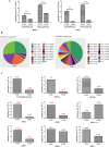
Figure 1
Comparative fidelity of plaque-purified viral populations and wild-type virus (NJ-Rc). Samples of the plaque-purified populations (3–2, 1–2, 18–1 and NJ-Rb) and NJ-Rc were cultured for 48 h in MARC-145 cells (MOI, 0.5) in the presence or absence of 0.5 mM guanidine (Gua). We compared the selected clones with or without 0.5 mM guanidine and found one clone, designated NJ-Rb, had the highest sensitivity to guanidine and the largest decrease in viral titre from 108 ± 0.15 TCID50/mL to 105.64 ± 0.13 TCID50/mL. A The resulting viral titres were determined using a TCID50 assay on MARC-145 cells. The experiment was performed in triplicate, and each error bar indicates one standard error of the mean (1 SEM). B The guanidine resistance ratios of the plaque-purified populations, NJ-Rb and NJ-Rc, were calculated as the viral titre in the presence of guanidine divided by the titre in the absence of guanidine.
Figure 2
Figure 2
Comparative mutation frequencies of PRRSV NJ-Rb and NJ-Rc (wild-type). A Mutation frequencies in the ORF5 and NSP2 regions of plaque-cloned, ribavirin-resistant mutant viruses (PRRSV NJ-Rb) after 10 passages in MARC-145 cells. Statistical significance was evaluated by determining the P-values. ***P < 0.001. B Abundance of quasispecies of NJ-Rb and NJ-Rc. The GeneID indicates the quasispecies ID, along with its sequence number (e.g., HAP0 ~ 19) and abundance information (e.g., 0.272952). The top 20 most abundant quasispecies were counted, and the rest were categorised as “Other”. The cumulative abundance of NJ-Rb quasispecies was more than 99.99%, and that of NJ quasispecies was more than 99.63%. (C) Full-length genome and ORF mutation frequency analysis. The red numbers represent the value. Except for ORF4 (red arrow), the mutation frequencies of the segmental genes in NJ-Rb, ORF1a, ORF1b, ORF2, ORF3, ORF5, ORF6 and ORF7 were significantly decreased.
Figure 3
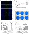
Figure 3
Rescue, biological characterisation and genetic stability of the reconstituted viruses. A Representative confocal images showing reconstituted viruses infecting MARC-145 cells. Forty-eight h post-transfection of MARC-145 cells with recombinant full-length virus clones, the cell monolayers were immunostained by an immunofluorescence assay. PRRSV was labelled with the N protein primary antibody (green), and nuclei were stained with DAPI (blue). B Multiple-step growth curves of viruses. The P2 reconstituted viruses vI218V, vS386P, vVP and vNJ-Rb, and the parental virus NJ-Rb, were used to infect fresh MARC-145 cells at an MOI of 0.1. The culture supernatants were collected and titrated at the indicated time points post-infection (pi). C Plaque morphology of viruses. At 4 dpi with the P2 reconstituted viruses (vI218V, vS386P, vVP, vNJ-Rb) and NJ-Rb, the MARC-145 cells were fixed, and plaques were visualised by crystal violet staining. Graphical representation of the mutation ORF5 (D) and NSP2 (E) frequencies of the reconstituted viruses. Statistical significance was evaluated by determining the P-values. *P < 0.05; **P < 0.01, ***P < 0.001.
Figure 4
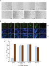
Figure 4
Observation of the reconstituted viruses and NJ-Rb in viral tissue tropism. A Growth characteristics of the reconstituted viruses (P1), NJ-Rb, NJ-Rc and NJ-a P80 in PAM cells, as observed by microscopy (100 ×). A viral CPE was observed in MARC-145 cells following NJ-Rc and NJ-a P80 infection. B Representative fluorescent images showing reconstituted viruses infecting CD163-transfected immortalised PAM cells (3D4/21). The viruses (vI218V, vS386P vVP, vNJ-Rb, NJ-Rb and NJ-Rc) were all viable as evidenced by IFA. PRRSV was labelled with the N protein primary antibody (green), and nuclei were stained with DAPI (blue). C Detection of residual virus loads in different tissues in groups challenged with each virus (NJ-Rb and NJ-Rc) via qRT-PCR (TaqMan chemistry). The residual virus loads were measured, and virus titres in different tissues were calculated and expressed as virus titres in TCID50/g equivalents based on the standard curve of the cycle threshold (CT) number plotted against the known virus titre of NJ-a p80. Asterisks indicate significant differences in residual virus loads in tissues compared to those of the NJ-Rc group (***P < 0.001).
Figure 5
Figure 5
Ribavirin-resistance profiles of the reconstituted viruses. A Representative images showing reconstituted virus infection in MARC-145 cells in the presence ( +) or absence (−) of 200 μM ribavirin (100 ×). A viral CPE was observed in MARC-145 cells. B TCID50 assay of the reconstituted viruses with or without ribavirin. Statistical significance was evaluated by determining the P-values. ***P < 0.001.
Figure 6
Figure 6
Construction and evaluation of the recombinant virus V218I and P386S based on NJ-a P80. A Multiple-step growth curves of viruses. The P2 reconstituted viruses vV218I, vP386S, vIS, vNJ-a P80, and the parental virus NJ-a P80 were used to infect fresh MARC-145 cells at an MOI of 0.1. The culture supernatants were collected and titrated at the indicated time points post-infection (pi). B TCID50 assay of the reconstituted viruses with or without ribavirin. C Graphical representation of the mutation ORF5 (C) and NSP2 (D) frequencies of the reconstituted viruses. Statistical significance was evaluated by determining the P-values. ***P < 0.001.
Figure 7
Figure 7
Sequence analysis of the NSP9 of PRRSVs. Sequence alignment of selected type 1 and type 2 PRRSV strains. The boxes show amino acids at residues 218 and 386. The red arrows show amino acids at residues 286 and 424. Dots represent identical amino acids with consensus sequences. The alignments were generated using the Cluster W method in the MEGALIGN software (DNASTAR Lasergene).
Figure 8
Figure 8
Predicted model of NSP9 and structure-based modulation of viral RdRp fidelity and ribavirin resistance. A Domain structure of NSP9 (RdRp). B Three views of the NSP9 structure generated by 90° rotations (left, back view; middle, side view; right, top view). C Aligned cartoon figure of PRRSV NSP9 and SARS-CoV-2 NSP12. The NSP12 is coloured in deep teal. D The positions of V218I and P386S (blue) and A286 and H424 (green-cyan), in the structural model. E Magnified cartoon figure of V218I. F Magnified cartoon figure of P386S. Colour code for NSP9 (NiRAN, interface, fingers, palm, and thumb) used throughout.

Similar articles

References

    1. Bloemraad M, de Kluijver EP, Petersen A, Burkhardt GE, Wensvoort G (1994) Porcine reproductive and respiratory syndrome: temperature and pH stability of Lelystad virus and its survival in tissue specimens from viraemic pigs. Vet Microbiol 42:361–371 - PubMed
    1. Lalonde C, Provost C, Gagnon CA (2020) Whole-genome sequencing of porcine reproductive and respiratory syndrome virus from field clinical samples improves the genomic surveillance of the virus. J Clin Microbiol 58:e00097-20 - PMC - PubMed
    1. Holtkamp DJ, Kliebenstein JB, Neumann EJ, Zimmerman JJ, Rotto HF, Yoder TK, Wang C, Yeske PE, Mowrer CL, Haley CA (2013) Assessment of the economic impact of porcine reproductive and respiratory syndrome virus on United States pork producers. J Swine Health Prod 21:72–84
    1. Hanada K, Suzuki Y, Nakane T, Hirose O, Gojobori T (2005) The origin and evolution of porcine reproductive and respiratory syndrome viruses. Mol Biol Evol 22:1024–1031 - PMC - PubMed
    1. Xing J, Zheng Z, Cao X, Wang Z, Xu Z, Gao H, Liu J, Xu S, Lin J, Chen S, Wang H, Zhang G, Sun Y (2022) Whole genome sequencing of clinical specimens reveals the genomic diversity of porcine reproductive and respiratory syndrome viruses emerging in China. Transbound Emerg Dis 69:e2530–e2540 - PubMed

MeSH terms

LinkOut - more resources